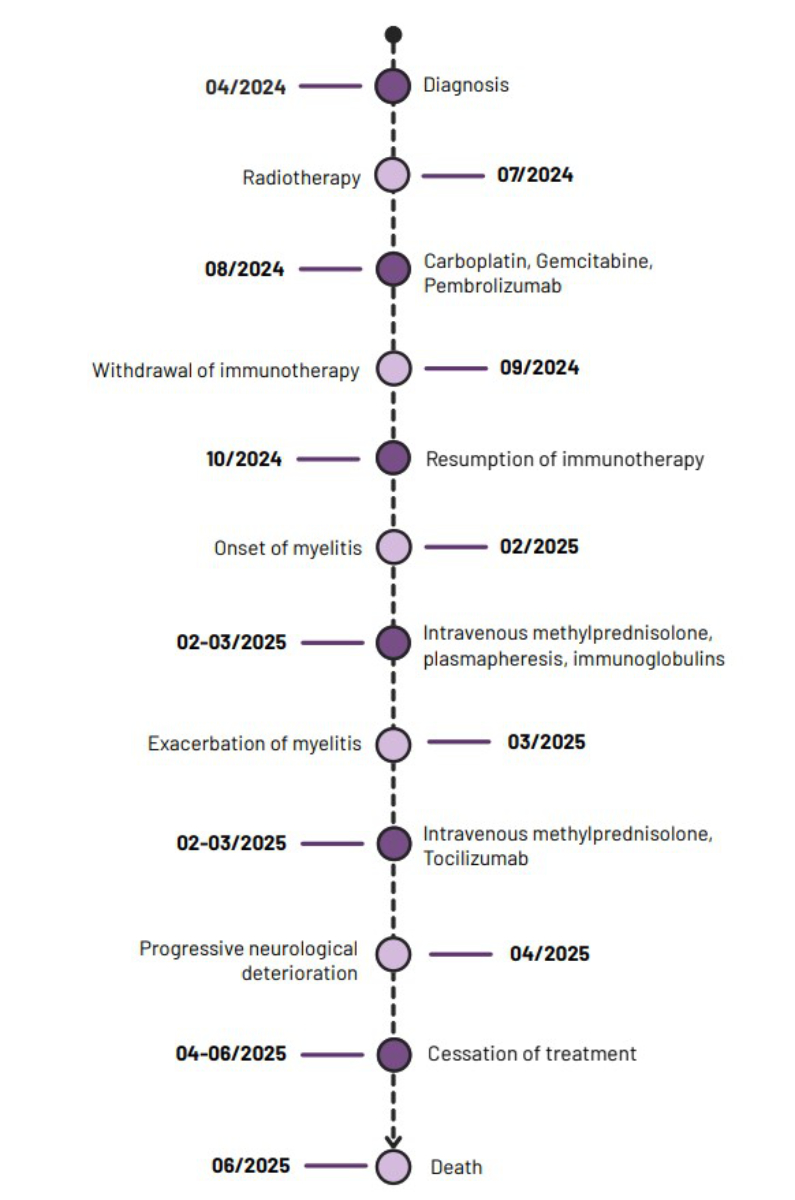
Figure 1Timeline of the patient’s events.
DOI: https://doi.org/https://doi.org/10.57187/5378
Immunotherapy with immune checkpoint inhibitors, such as pembrolizumab, has profoundly transformed the management of non-small-cell lung cancer (NSCLC), particularly in advanced or metastatic stages [1]. By blocking the interaction between programmed cell death protein 1 (PD-1) and its ligand PD-L1, these treatments restore cytotoxic T-cell activity and may lead to durable antitumour responses [1]. However, immune activation induced by immune checkpoint inhibitors can lead to immune-related adverse events involving multiple organs, including the nervous system [2, 3]. Neurological complications are rare but may result in significant functional impairment and can be life-threatening. Among these, inflammatory myelitis represents a rare yet particularly severe complication, with complex diagnostic and therapeutic challenges [2, 4].
The pathophysiology of immune checkpoint inhibitor-associated myelitis remains poorly understood and diagnosis is primarily based on the exclusion of alternative diagnoses supported by clinico-radiological concordance [4]. Aggravating or predisposing factors have been suggested, notably prior exposure to radiotherapy involving radiosensitive neurological structures such as the spinal cord. Here we report a case of extensive cervical myelitis occurring after pembrolizumab treatment in a patient previously treated with cervical radiotherapy, illustrating the diagnostic difficulties and raising the question of a potential synergistic neurotoxicity between immunotherapy and radiotherapy.
Given the descriptive nature of a single-patient case report, no statistical analyses were performed.
In early 2024, a patient in his 70s with a medical history notable for chronic kidney disease (KDIGO IV), ischaemic stroke in 2018, type 2 diabetes mellitus and structural epilepsy was evaluated with progressively worsening chronic cervical pain. Cervical magnetic resonance imaging (MRI) performed in April 2024 revealed a lesion involving a secondary malignant lesion in the posterior arch of C3 without spinal cord compression. Further investigations identified a right upper-lobe lung adenocarcinoma with bone (C3, right first rib), pulmonary, lymph node and renal metastases, classified as T2N1M1c (stage IV disease according to the UICC classification).
Immunohistochemical analysis revealed low PD-L1 expression (<1%), TTF-1 positivity, HER2 and MET co-expression, with no EGFR, KRAS, ALK, ROS1, BRAF or MET exon 14 skipping mutations. Next-generation sequencing identified a TP53 mutation (A159V).
The patient received highly targeted cervical radiotherapy to the C3 vertebral lesion, with no significant irradiation of other spinal cord segments beyond minimal scatter exposure (with 30 Gy in 5 fractions, Dmax spinal cord 24.7 Gy) in July 2024, followed by combined immunochemotherapy with carboplatin, gemcitabine and pembrolizumab initiated in August 2024. Treatment was interrupted after two cycles due to severe acute pyelonephritis complicated by acute renal failure requiring ureteral stenting. Pembrolizumab was resumed in October 2024. A positron emission tomography-computed tomography (PET-CT) scan in November 2024 demonstrated partial tumour response and partial remission of the cervical metastasis.
In February 2025, the patient was admitted for rapidly progressive right-sided hemiparesis developing over several days. Neurological examination revealed spasticity with moderate-to-severe weakness of the right upper and lower limbs with a severe afferent ataxia and severe fine motor skill impairment of the right hand. A brain MRI ruled out ischaemic or haemorrhagic stroke as well as metastatic disease. Electroencephalography showed no seizures. The MRI of the cervical spine revealed an extensive intramedullary contrast-enhancing T2 hyperintensity at level C3 to C5 with oedematous swelling of the myelon extending from the brain stem to T4 consistent with inflammatory cervical myelitis.
In light of the rapid neurological deterioration with the development of asymmetric tetraparesis predominantly affecting the right side, lumbar puncture was performed. Cerebrospinal fluid analysis revealed moderate pleocytosis (10 cells/mcl), markedly elevated cerebrospinal fluid protein level (3200 mg/l) and an increased cerebrospinal fluid-to-serum albumin ratio, consistent with blood–brain barrier disruption. Cytological examination showed no malignant cells. Cerebrospinal fluid molecular analysis did not identify any pathogenic mutations, including the TP53 variant previously detected on the initial tissue biopsy. Extensive infectious investigations, including serologies and PCR testing of cerebrospinal fluid (HIV, syphilis, Borrelia, multiplex PCR) were negative, and no evidence of infection with myotropic pathogens, including Coxsackieviruses, was identified; the workup for autoimmune encephalitis/myelitis (including anti-aquaporin antibodies and the anti-MOG antibodies) was also negative.
The close temporal relationship between the onset of neurological symptoms during pembrolizumab treatment, the absence of infectious, neoplastic or alternative aetiologies, and the findings from spinal MRI and cerebrospinal fluid analysis led to the diagnosis of pembrolizumab-induced inflammatory myelitis.
The patient’s clinical course is summarised in a timeline (figure 1).
Figure 1Timeline of the patient’s events.
Pembrolizumab was discontinued after nine cycles. Treatment consisted of high-dose intravenous methylprednisolone (1 g/day for 3 days), followed by five sessions of plasmapheresis and intravenous immunoglobulin (Privigen 35 g/day), which were discontinued on day 4 due to a severe cutaneous allergic reaction. Partial motor improvement was observed; however, persistent disability required wheelchair use.
In March 2025, the neurological status deteriorated again, with rapidly progressive flaccid paraplegia predominantly affecting the right side, associated with a thoracic sensory level and acute bladder and sphincter dysfunction. Repeat spinal MRI demonstrated persistent extensive cervical intramedullary T2 hyperintensity extending to the T4 level (figure 2). A second course of high-dose methylprednisolone was administered, followed by oral tapering and initiation of tocilizumab (anti-interleukin-6 [IL-6]) at a dose of 50 mg administered every four weeks. Symptomatic antispastic treatment was also initiated.
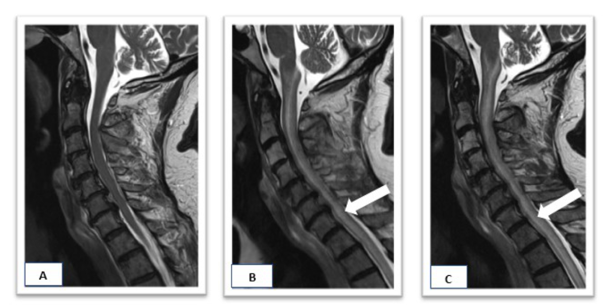
Figure 2Comparative spinal MRI showing the initial examination in April 2024 (A), MRI at the time of myelitis diagnosis (white arrow) in February 2025 (B) and radiological progression of myelitis (white arrow) in March 2025 (C).
Despite aggressive immunosuppressive therapy and rehabilitation, the neurological outcome remained unfavourable. The patient was transferred to palliative care in June 2025 and died a few days after.
This case illustrates an extensive cervical myelitis associated with pembrolizumab, characterised by a rapidly progressive course and an insufficient response to conventional immunosuppressive therapies. Immune checkpoint inhibitor-induced myelitis is generally reported to respond favourably to early initiation of high-dose corticosteroid therapy, sometimes in combination with second-line therapies such as plasmapheresis or intravenous immunoglobulin [2]. In contrast, the clinical course observed in our patient highlights a particularly severe and refractory form, with persistent functional impairment despite aggressive management. This observation underscores the heterogeneity of the clinical spectrum of immune-mediated myelitis and the substantial challenges associated with the management of extensive spinal cord involvement, particularly in the presence of potentially aggravating factors such as prior cervical radiotherapy.
Neurological immune-related adverse events associated with immune checkpoint inhibitors are rare but potentially life-threatening, accounting for approximately 1% of all immune-related adverse events [5, 6]. The overall incidence of neurological immune-mediated adverse events of any grade is estimated at 6.1% in patients treated with anti-PD-1 agents [2], whereas severe forms (grade ≥3) occur in fewer than 1% of cases [2, 3, 6]. Among these, inflammatory transverse myelitis represents a particularly uncommon entity, most often presenting as a longitudinally extensive spinal cord lesion involving multiple segments and associated with an unfavourable functional prognosis [5]. Clinically, these forms typically manifest with rapidly progressive motor, sensory and/or sphincter deficits, often leading to significant disability and requiring urgent intervention [3, 5–7].
In the literature, pembrolizumab-induced myelitis typically occurs after several treatment cycles, with a highly variable delay ranging from a few weeks to several months after treatment initiation [3–5, 7, 8]. Reported imaging findings frequently describe extensive intramedullary T2 hyperintense lesions on MRI, often without contrast enhancement [3, 9].
Histopathological data regarding pembrolizumab-induced myelitis remain extremely limited, as spinal cord biopsy or autopsy is rarely performed. The few available pathological reports suggest a predominantly T-cell-mediated inflammatory process [3, 9]. Cerebrospinal fluid abnormalities are generally nonspecific and typically include lymphocytic pleocytosis, elevated protein levels, normal glucose concentration and negative cytology, as observed in our patient [3, 9].
In our case, extensive investigations excluded infectious, neoplastic and primary autoimmune aetiologies. Although creatine kinase levels were not measured, the absence of myalgia or clinical signs of myositis made inflammatory or statin-associated myopathy unlikely, particularly as statin therapy had already been discontinued at admission.
Furthermore, the causal relationship between pembrolizumab and the occurrence of myelitis in our patient is supported by several elements consistent with Bradford Hill’s criteria for causality, including the temporal association between drug exposure and symptom onset, the exclusion of alternative aetiologies and the concordance with previously reported cases in the literature [10]. These findings further support the diagnosis of an immune-mediated neurological adverse event associated with immune checkpoint inhibition [2].
A notable feature of this case is the history of prior cervical radiotherapy, raising the question of a “radiation recall myelitis” phenomenon or a synergistic inflammatory toxicity between radiotherapy and immunotherapy. Radiation recall phenomena typically occur weeks to months after exposure to ionising radiation and may present with local neurological deficits [8, 11]. In our patient, the administered radiation dose of the spinal cord was 24.7 Gy in 5 fractions (EQD2 <40 Gy for α/β 3 Gy) and thus below the classic thresholds associated with radiation-induced myelopathy, which are usually reported for cumulative doses exceeding 45–50 Gy (EQD2 for α/β 3 Gy) [8]. However, rare cases of myelitis occurring at lower doses have been described, particularly in the context of concomitant immune activation, suggesting an immune-exacerbated mechanism rather than isolated radiation injury [8, 11, 12]. Furthermore, the cervical spinal cord is recognised as a particularly vulnerable neurological structure, and the delayed onset of symptoms following irradiation is compatible with a “radiation recall myelitis” [13]. Although the most pronounced inflammatory changes were observed at the C3–C5 levels corresponding to the region surrounding the previously irradiated C3 lesion, the oedema extended beyond this region to the thoracic spinal cord, suggesting a more diffuse inflammatory process rather than a purely localised radiation recall phenomenon. Nevertheless, the predominance of abnormalities within the irradiated region supports a potential role of prior radiotherapy as a local susceptibility factor. The association of immunotherapy and radiotherapy in such radiosensitive regions therefore warrants heightened vigilance, as these treatment modalities may interact synergistically, increasing the risk and severity of neurological toxicity [11, 12].
Distinguishing between a purely immune-mediated myelitis induced by immune checkpoint inhibitors and a radiation-induced myelitis exacerbated by immune activation remains challenging but carries important therapeutic implications. In our case, lesion topography, symptom chronology and poor response to immunosuppressive therapies support the hypothesis of a mixed pathophysiological mechanism. In this context, early multidisciplinary management and close neurological monitoring are essential to limit long-term functional sequelae [8, 11, 12].
To better contextualise our observation, we compared reported cases of immune checkpoint inhibitor-induced myelitis according to prior spinal radiotherapy exposure and neurological outcomes (table 1). Although prior radiotherapy was not uniformly associated with poorer prognosis, severe or refractory presentations appeared proportionally more frequent among previously irradiated patients. In this context, the rapidly progressive and fatal course observed in our patient, despite early escalation to multimodal immunosuppressive therapy, supports the hypothesis that prior spinal irradiation may act as a vulnerability factor, potentially amplifying immune-mediated inflammatory injury under checkpoint inhibition. Given the limited number and heterogeneity of available reports, these observations should be interpreted cautiously and warrant further investigation in larger datasets.
Table 1Reported cases of immune checkpoint inhibitor-induced myelitis according to prior spinal radiotherapy exposure and clinical outcomes.
| Author (year) | ICI class | Prior spinal radiotherapy | Escalation therapy | Outcomes |
| Charabi et al. (2021) [3] | PD-1 | No | Steroids + IVIG + PLEX | Death |
| Gritsch et al. (2022) [4] | PD-1 | No | Steroids | Partial remission |
| Chatterton et al. (2023) [5] | PD-1 | No | Steroids + IVIG | Complete remission |
| PD-1 | No | Steroids | Complete remission | |
| PD-1 + CTLA-4 | Yes | Steroids + IVIG + PLEX | Refractory | |
| PD-1 | Yes | Steroids +IVIG | Death | |
| Pietak et al. (2025) [7] | PD-1 + ITK | Yes | Steroids + IVIG | Partial remission |
| Owen et al. (2022) [8] | PD-1 | Yes | Steroids + IVIG + second-line immunosuppression* | Partial remission |
| Vickers et al. (2020) [9] | PD-1 | No | Steroids + IVIG | Partial remission |
| PD-1 | No | Steroids + PLEX | Partial remission | |
| PD-1 | No | Steroids + IVIG + PLEX | Complete remission | |
| Carausu et al. (2019) [11] | PD-1 | Yes | Steroids | Complete remission |
| Chang et al. (2018)[12] | PD-1 + CTLA-4 | Yes | Steroids + IVIG + PLEX + second-line immunosuppression ** | Death |
| Picca et al. (2021) [14] | PD-1 + CTLA-4 | No | Steroids + PLEX + second-line immunosuppression *** | Refractory |
| Our case (2026) | PD-1 | Yes | Steroids + IVIG + PLEX + second- line immunosuppression **** | Death |
* mycophenolate mofetil
** cyclophosphamide + infliximab
*** cyclophosphamide + tocilizumab
**** tocilizumab
Abbreviations: CTLA-4: cytotoxic T-lymphocyte antigen-4; ITK: tyrosine kinase inhibitor; ICI: immune checkpoint inhibitor; IVIG: intravenous immunoglobulin; PD-1: programmed cell death protein 1; PLEX: plasmapheresis.
Regarding therapeutic management, high-dose intravenous corticosteroids remain the recommended first-line treatment following permanent discontinuation of immune checkpoint inhibitors. In cases of insufficient response, plasmapheresis and intravenous immunoglobulin are proposed as second-line options [2, 4, 7, 8, 12]. A case published in 2022 reported significant clinical improvement following combined corticosteroid and intravenous immunoglobulin therapy in a patient with metastatic NSCLC treated with pembrolizumab and palliative radiotherapy, highlighting the potential benefit of a combined approach in severe cases [8]. In refractory cases, additional immunomodulatory strategies targeting pro-inflammatory cytokines may be considered [14]. Tocilizumab was introduced as a third-line therapy in our patient because of its ability to inhibit the IL-6 pathway, which is increasingly implicated in immune-mediated neurotoxicity [14]. IL-6 blockade is now widely used in the management of severe neuroinflammatory toxicities, particularly in patients receiving bispecific antibodies or CAR T-cell therapies [15]. Although IL-6 levels were not measured in our patient, elevated cytokine levels have been reported in similar contexts, supporting the potential role of tocilizumab in refractory immune-mediated neurological adverse events. Other immunosuppressive agents, including rituximab, infliximab, cyclophosphamide or extracorporeal photopheresis, have been reported in isolated cases of refractory immune checkpoint inhibitor-induced myelitis, although current evidence remains insufficient to support their systematic use [12]. Despite transient improvement under intensive immunosuppressive therapy, the neurological outcome in our patient remained unfavourable, illustrating the potentially devastating nature of these complications.
The comprehensive exclusion of competing diagnoses and the strong clinicoradiological correlation further strengthen the robustness of our case. However, several limitations should be acknowledged. Given the single-case nature of this report, caution is required when extrapolating these findings. In addition, the absence of histopathological confirmation and detailed immunological analyses limits pathophysiological interpretation. Finally, prior cervical radiotherapy represents a potential confounding factor, making it difficult to distinguish between a primary immune-mediated adverse event, a radiation recall phenomenon or a combination of both.
This case highlights several key messages for clinical practice. First, any new neurological symptom in a patient receiving immune checkpoint inhibitors should prompt rapid neurological and radiological evaluation, particularly in the presence of prior radiotherapy involving sensitive structures such as the spinal cord. Second, early multidisciplinary management involving oncologists, neurologists and radiation oncologists is essential to optimise functional prognosis.
This case highlights a rare but severe neurological complication of pembrolizumab immunotherapy. A potential exacerbation by prior cervical radiotherapy cannot be completely excluded, but the treated volume in this case was significantly smaller than the affected myelon. Distinguishing between a purely immune-mediated toxicity and a radiation-induced myelitis exacerbated by immune activation remains a significant challenge with important therapeutic implications. The combination of corticosteroids, plasma exchange, intravenous immunoglobulin and IL-6 inhibition with tocilizumab may allow partial control of inflammation, although prognosis remains poor in severe and refractory forms. Finally, this observation underscores the need for close neurological monitoring when immune checkpoint inhibitors are administered in combination with radiotherapy involving radiosensitive structures such as the spinal cord, in order to enable early and appropriate management.
Informed consent: As this case report was written after the patient’s death, consent for publication was obtained from the patient’s wife and a formal waiver of medical confidentiality was granted by the Cantonal Medical Officer of the Canton of Jura.
The authors did not receive any financial support for the preparation of this manuscript.
All authors have completed and submitted the International Committee of Medical Journal Editors form for disclosure of potential conflicts of interest. No potential conflict of interest related to the content of this manuscript was disclosed.
1. Kwok G, Yau TC, Chiu JW, Tse E, Kwong YL. Pembrolizumab (Keytruda). Hum Vaccin Immunother. 2016 Nov;12(11):2777–89.
2. Anderson D, Beecher G, Nathoo N, Smylie M, McCombe JA, Walker J, et al. Proposed diagnostic and treatment paradigm for high-grade neurological complications of immune checkpoint inhibitors. Neurooncol Pract. 2019 Sep;6(5):340–5.
3. Charabi S, Engell-Noerregaard L, Nilsson AC, Stenör C. Longitudinal extensive transverse myelitis with novel autoantibodies following two rounds of pembrolizumab. Front Neurol. 2021 Apr;12:655283.
4. Gritsch D, Valencia-Sanchez C. Drug-related immune-mediated myelopathies. Front Neurol. 2022 Sep;13:1003270.
5. Chatterton S, Xi S, Jia JX, Krause M, Long GV, Atkinson V, et al. Case series: immune checkpoint inhibitor-induced transverse myelitis. Front Neurol. 2023 Feb;14:1130313.
6. Payá PR, Abad ÁM, Mirete BG, Arias AF, Rivilla MB, Benítez Fuentes JD, et al. Successful management of immune-mediated myelitis in a renal cancer patient: a case report. Case Rep Oncol. 2025 Jun;18(1):892–9.
7. Piętak M, Kruczyk B, Domański P, Jarosińska J, Hołdakowska A, Demkow T, et al. Longitudinally extensive transverse myelitis after pembrolizumab and lenvatinib therapy for a rare subtype of renal cell carcinoma: A case report. Oncol Lett. 2025 Jun;30(2):386.
8. Owen T, Fung AS. Combination intravenous immune globulin (IVIG) and high-dose steroids for treatment of immune-related myelitis in a non-small cell lung cancer patient treated with pembrolizumab and palliative radiation : a case report. Clin Lung Cancer. 2022 Dec;23(8):e563–7.
9. Vickers ML, Seidl B, Bigby K, Chern B, Eriksson L, Hartnett G, et al. Inflammatory myeloradiculitis secondary to pembrolizumab: a case report and literature review. Case Rep Oncol Med. 2020 Aug;2020:8819296.
10. Hill AB. The Environment and Disease: association or Causation? Proc R Soc Med. 1965 May;58(5):295–300.
11. Carausu M, Beddok A, Langer A, Girard N, Bidard FC, Massiani MA, et al. Radiation myelitis after pembrolizumab administration, with favorable clinical evolution and safe rechallenge: a case report and review of the literature. J Immunother Cancer. 2019 Nov;7(1):317.
12. Chang VA, Simpson DR, Daniels GA, Piccioni DE. Infliximab for treatment-refractory transverse myelitis following immune therapy and radiation. J Immunother Cancer. 2018 Dec;6(1):153.
13. Schultheiss TE, Kun LE, Ang KK, Stephens LC. Radiation response of the central nervous system. Int J Radiat Oncol Biol Phys. 1995 Mar;31(5):1093–112.
14. Picca A, Valyraki N, Birzu C, Kramkimel N, Hermine O, Zahr N, et al. Anti–interleukin-6 and Janus kinase inhibitors for severe neurologic toxicity of checkpoint inhibitors. Neurol Neuroimmunol Neuroinflamm. 2021 Sep;8(6):e1073.
15. Brudno JN, Maus MV, Hinrichs CS. CAR T Cells and T-Cell Therapies for Cancer: A Translational Science Review. JAMA. 2024 Dec;332(22):1924–35.