Granulomatous tubulointerstitial nephritis secondary to small lymphocytic
lymphoma: a case report
DOI: https://doi.org/https://doi.org/10.57187/5364
Ianis Petignata,
Aurélie Huberb,
Diogo Da Silvaa,
Carmen de Ramon Ortizc,
Grégoire Arnouxd,
Sophie de Seigneuxb,
Lena Berchtoldb
a General Internal Medicine Service,
Department of Medicine, Geneva University Hospitals, Geneva, Switzerland
b Nephrology
and Hypertension Service, Department of Medicine, Geneva University Hospitals,
Geneva, Switzerland
c Hematology Service, Department of Oncology, Geneva
University Hospitals, Geneva, Switzerland
d Clinical Pathology
Service, Diagnostic Department, Geneva University Hospitals, Geneva,
Switzerland
Summary
BACKGROUND: Granulomatous tubulointerstitial nephritis is an uncommon
cause of acute kidney injury with a broad differential diagnosis that includes
drug exposure, infections, autoimmune conditions, and sarcoidosis. Although
renal infiltration by small lymphocytic lymphoma/chronic lymphocytic leukaemia
(SLL/CLL) is frequently observed in autopsy series, it rarely results in
clinically significant renal dysfunction. Granulomatous interstitial nephritis
directly related to lymphomatous infiltration is particularly rare, with only a
few cases described in the literature.
CASE PRESENTATION: A man in his seventh decade with indolent SLL developed rapidly
progressive acute kidney injury. Extensive evaluation excluded obstructive,
infectious (including tuberculosis), autoimmune, and drug-related causes, and sarcoidosis
was considered unlikely in the absence of supportive clinical and laboratory
findings. Immunohistochemistry and molecular techniques demonstrated a clonal
B-cell population consistent with SLL, establishing the diagnosis of
lymphoma-related granulomatous tubulointerstitial nephritis. Treatment with high-dose
corticosteroids and
acalabrutinib improved renal function, though targeted therapy was discontinued
due to severe infectious complications.
CONCLUSION: Granulomatous tubulointerstitial nephritis secondary to lymphomatous infiltration
should be considered in
the differential diagnosis of unexplained acute kidney injury in patients with indolent
CLL/SLL. Immunohistochemistry is crucial for confirming B-cell infiltration.
Corticosteroids and targeted therapies may lead to renal improvement but
require careful monitoring given the risk of infectious complications. This
case adds a rare and well-documented presentation of SLL-associated granulomatous
tubulointerstitial nephritis to the
current medical literature.
Introduction
Granulomatous tubulointerstitial
nephritis is a rare renal condition, observed in approximately 0.3% to
0.9% of kidney biopsies. It is histologically defined by interstitial
inflammation and the presence of granulomas. Renal involvement may be isolated
or occur in the context of a systemic disease, which can help guide the
diagnostic evaluation [1].
The aetiologies of granulomatous tubulointerstitial nephritis are diverse. Drug-induced
forms are frequently reported among secondary causes and are primarily associated
with non-steroidal anti-inflammatory drugs (NSAIDs), proton pump inhibitors
(PPIs), diuretics, certain antibiotics, antiepileptic drugs, and some immune
checkpoint inhibitors. Infectious causes, especially mycobacterial or fungal
infections, should also be considered, particularly in endemic areas and in
immunocompromised patients. Autoimmune and autoinflammatory diseases are other recognised
aetiologies, including sarcoidosis, systemic lupus erythematosus,
antineutrophil cytoplasmic antibody (ANCA)-associated vasculitis,
tubulointerstitial nephritis with uveitis (TINU), and chronic inflammatory
bowel diseases. Approximately 10% of granulomatous tubulointerstitial nephritis cases
are idiopathic [1].
Chronic lymphocytic leukaemia (CLL)
and small lymphocytic lymphoma (SLL) are mature B-cell malignancies characterised
by the clonal proliferation of B lymphocytes and typically follow an indolent
course. The term SLL is used when lymph node involvement is predominant without
significant lymphocytosis in the peripheral blood, whereas CLL refers to cases
with lymphocytosis, defined as a monoclonal B-cell count of at least 5 x 10⁹/l
in the peripheral blood. CLL and SLL are considered different clinical
manifestations of the same disease entity. CLL/SLL is the most common leukaemia
in adults in developed countries. It occurs more frequently in men (male-to-female
ratio of 1.2:1) and typically presents at a median age of approximately 70
years. Most patients are asymptomatic at diagnosis, which is often made
following the discovery of persistent lymphocytosis or, less commonly, cervical
lymphadenopathy during routine evaluation [2].
Although renal involvement by CLL/SLL
is common in autopsy series, reported in 60 to 90% of cases, it is typically
subclinical and rarely leads to significant impairment of renal function [3, 4]. Consequently,
CLL/SLL is rarely
considered in the differential diagnosis of acute kidney injury. Within the
spectrum of CLL-associated renal lesions, granulomatous tubulointerstitial nephritis
is particularly rare. In a
25-year multicentre series, only a small fraction of patients with
CLL/SLL-related kidney disease exhibited interstitial granulomas directly
attributable to the malignancy [5]. In this context, the diagnosis of
granulomatous tubulointerstitial nephritis as a direct manifestation of CLL/SLL represents
an exceptional and
diagnostically challenging clinical entity.
Here, we present a case of
granulomatous tubulointerstitial nephritis secondary to renal infiltration by
monoclonal B cells in a patient with SLL, along with a review of similar rare
cases described in the literature.
Case presentation
We report the case of a man in his
seventh decade with a history of Parkinson’s disease who was referred to a haematologist
in 2023 following the incidental discovery of isolated cervical
lymphadenopathy. At presentation, the patient reported weight loss without
fever or night sweats and had no other symptoms. Physical examination revealed
an extrapyramidal syndrome with bilateral cervical and axillary
lymphadenopathy, with no evidence of inguinal lymphadenopathy or
hepatosplenomegaly. The patient’s general condition was preserved. A lymph node
biopsy revealed clonal B-cell proliferation expressing CD5, CD19, CD22, CD23,
CD43, CD52, CD79b, and surface IgM, with the absence of CD10, CD11c, CD25,
CD38, CD103, or FMC7. Positron emission tomography-computed tomography (PET-CT)
total body imaging showed additional lymphadenopathy in the cervical,
mediastinal, and retroperitoneal regions, supporting the diagnosis of SLL. In
the absence of lymphocytosis, clinical symptoms, or evidence of disease
progression, close haematological surveillance was implemented without
indication for specific therapy.
From early 2024, a gradual increase in
serum creatinine was observed, rising from a baseline of 100 µmol/l to 196
µmol/l by October. The patient was referred to the nephrology outpatient
clinic. To rule out ureteral obstruction secondary to retroperitoneal
lymphadenopathy, MAG3 renal scintigraphy with a diuretic challenge was
performed. The scan demonstrated bilateral impaired renal excretion, more
pronounced on the right side.
In January 2025, the patient was
admitted for rapidly progressive acute kidney injury, with serum creatinine
reaching 558 µmol/l. At admission, the patient reported only difficulty
initiating voiding, which had been present for 2 months. He did not report
oliguria, dysuria, or haematuria. He also denied asthenia, recent weight loss,
or night sweats. Physical examination revealed supradiaphragmatic
lymphadenopathy involving the cervical, supraclavicular, and axillary regions,
without evidence of enlargement according to the haematologist following the
patient in the outpatient setting. The remainder of the physical examination was
unremarkable. Emergency placement of a right pigtail catheter did not improve
renal function. A laboratory workup showed no systemic inflammation (CRP 4.45
mg/l) or electrolyte abnormalities. Urinalysis was unremarkable, without
leukocyturia, and showed hyaline casts (++). Twenty-four-hour proteinuria was
moderate (0.8 g/24 h), with a predominance of non-albumin proteins, suggesting a
non-glomerular pattern of protein loss. Kidney biopsy revealed diffuse and
severe tubulointerstitial nephritis with an abundant mononuclear cell-rich
infiltrate and non-necrotising granulomas with multinucleated giant cells (figure
1).
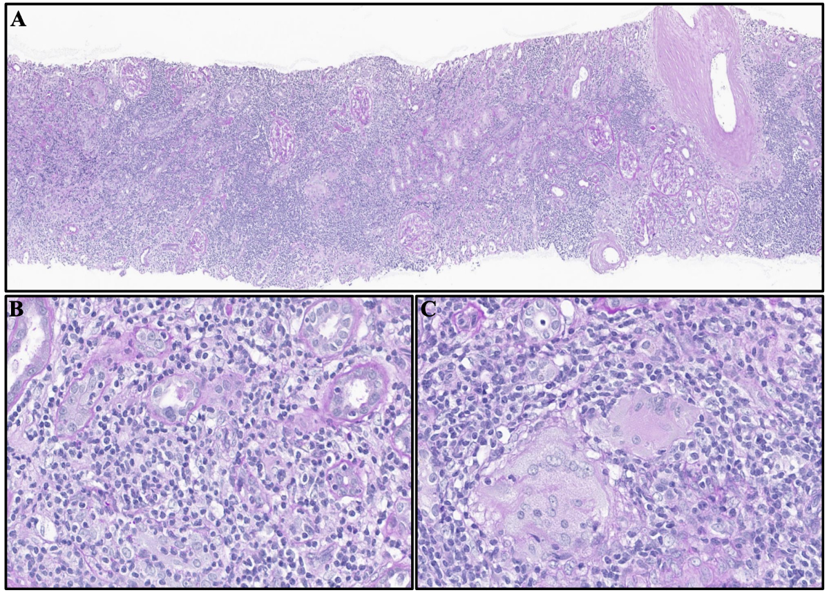
Figure 1A. Light microscopy revealed a
severe and diffuse cellular interstitial infiltrate, with multiple non-necrotising
granulomas. Haematoxylin-eosin/Jones stain. Formalin-fixed paraffin-embedded
section; 10× magnification. B and C. The interstitial infiltrate
consisted mainly of non-atypical mononuclear cells and a few poorly organised
non-necrotising epithelioid granulomas with multinucleated giant cells (C).
Haematoxylin-eosin stain. Formalin-fixed paraffin-embedded section; 40×
magnification.
A thorough aetiological workup was
undertaken. There was no recent change in the patient’s usual medication and no
exposure to medication known to induce interstitial nephritis. He was receiving
calcium, vitamin D, vitamin B12, and folic acid supplementation, rosuvastatin,
and, for Parkinson’s disease, opicapone, pramipexole, and a combination of
levodopa and benserazide. The patient had no risk factors for tuberculosis, no
clinical or radiological signs of mycobacterial infection, and negative TB-Spot
results. Polymerase chain reaction analysis performed on the kidney biopsy
found no evidence of Mycobacterium tuberculosis complex DNA. Autoimmune
evaluation was also non-contributory, with negative antinuclear antibodies and
ANCA, as well as normal C3/C4 complement levels. Serum immunofixation revealed
a small monoclonal IgG lambda peak, with a normal kappa/lambda free light chain
ratio. Quantitative immunoglobulin testing revealed decreased levels of IgG at
3.98 g/l (reference range 7–16 g/l), IgA at 0.30 g/l (reference range 0.7–4
g/l), and IgM at <0.05 g/l (reference range 0.4–2.3 g/l). Sarcoidosis was
deemed unlikely given the absence of hypercalcaemia, hypercalciuria, or
elevated 25-(OH) vitamin D (87 nmol/l) and angiotensin-converting enzyme (46
U/l) levels. 1,25-(OH)₂ vitamin D and sIL2 were not measured. The 18F-FDG
PET-CT showed no signs of hypermetabolic lymphadenopathy, which is typically seen
in sarcoidosis. PET-CT confirmed the presence of multiple non-hypermetabolic
lymphadenopathies (cervical, axillary, and infra-diaphragmatic), consistent
with SLL. Given the lack of alternative explanations, specific
immunohistochemical staining was performed on the renal biopsy. This confirmed
infiltration by CLL/SLL-type B-cell lymphoma with the following expression
profile: CD20 low, CD5 positive, CD23 positive, and BCL1. The inflammatory
infiltrate was heterogeneous, with a mixture of T cells (CD3-positive and CD5-positive)
and, to a lesser extent, B cells (CD20-positive and CD79-positive). The spatial
distribution appeared random rather than being localised to the periphery of
granulomas (figure 2). Next-generation sequencing (NGS) identified a mutation
in the SF3B1 gene, without a TP53 abnormality, strongly supporting the
probability of an SLL B-cell clone [6].
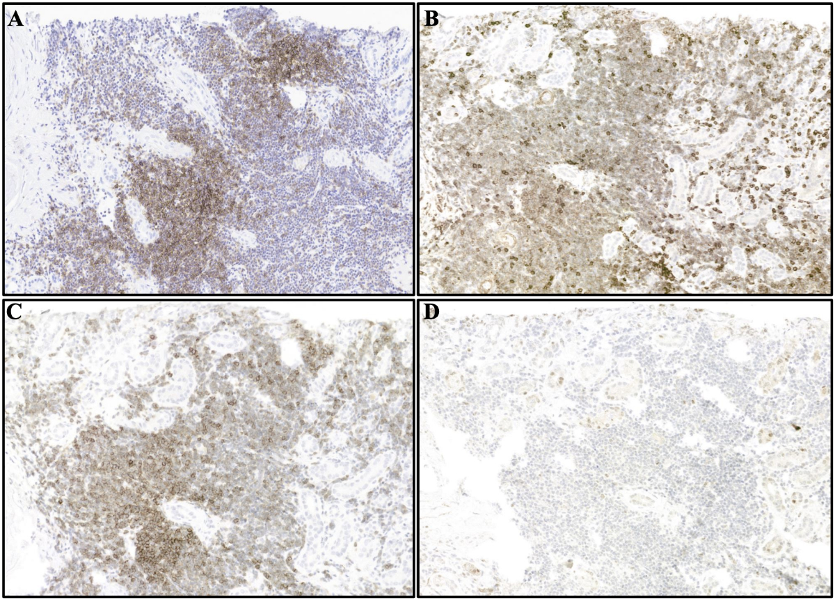
Figure 2Immunochemistry study revealed kidney
infiltration by small lymphocytes with a characteristic immunophenotype showing
diminished CD20 expression (A), aberrant CD5 expression (B), along with CD23
expression (C), and the absence of significant BCL1 staining (D), consistent
with small lymphocytic lymphoma. Formalin-fixed paraffin-embedded section with
DAB as chromogen; 20× magnification.
The diagnosis of granulomatous
tubulointerstitial nephritis secondary to renal infiltration by SLL was
established. Treatment was initiated with intravenous methylprednisolone (500
mg/day for three days), followed by oral prednisone at 1 mg/kg/day. In parallel,
and after multidisciplinary discussion with the haematology team, acalabrutinib,
an oral Bruton tyrosine kinase inhibitor, was started due to the lymphomatous
renal infiltration. The initial evolution was favourable, with progressive
improvement in renal function and stabilisation of serum creatinine at 190
µmol/l four months after treatment initiation and at the time of corticosteroid
tapering. However, acalabrutinib had to be discontinued early because of major
infectious complications that occurred in the weeks following its initiation.
The patient first developed influenza A pneumonia, which was rapidly
complicated by bacterial superinfection with an unidentified pathogen. A few
weeks later, he developed invasive fungal sinusitis due to Aspergillus
fumigatus, with secondary bony erosion resulting from the Aspergillus
infection, complicated by a right abducens nerve palsy in April 2025. The
patient underwent urgent sphenoidotomy due to cranial nerve involvement. Due to
osseous involvement, treatment with posaconazole and anidulafungin was
initiated and continued for two weeks, followed by therapy with inhaled
amphotericin B and oral posaconazole. One month later, the patient presented
with bacteraemia due to Staphylococcus epidermidis (3 out of 4 blood
culture sets) and Staphylococcus hominis (3 out of 4 blood culture
sets), secondary to bacterial sphenoidal sinusitis and a persistent bony defect
following invasive aspergillosis. Management required prolonged hospitalisation
and a one-week course of cefazolin, with a favourable clinical outcome. The
patient underwent serial sinus imaging follow-up after hospital discharge.
Given the favourable clinical and radiological evolution, inhaled amphotericin
B was continued for a total of three months, and posaconazole for ten months. Due
to the severity of the infectious complications, after further discussion with
the haematologists, acalabrutinib was suspended, and therapy with obinutuzumab,
a humanised type II anti-CD20 monoclonal antibody, and venetoclax, a selective
BCL-2 inhibitor, was planned. This therapy has not yet been started because of
the recent infections (timeline shown in figure 3).
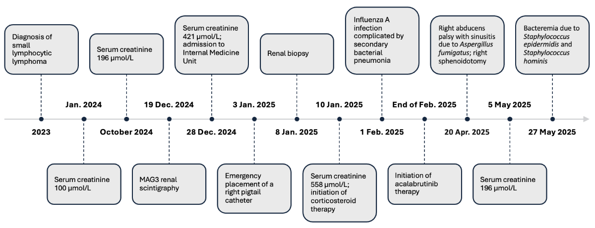
Figure 3Timeline from the diagnosis of small
lymphocytic lymphoma to the acute care episode.
Discussion
This
study reports a new case of granulomatous tubulointerstitial nephritis in a patient
with renal infiltration by SLL. In
patients with CLL/SLL, the most commonly reported causes of acute kidney injury
include acute tubular necrosis, uric acid crystal nephropathy, light chain
nephropathy, obstructive nephropathy, amyloidosis, hypercalcaemia, certain forms
of glomerulonephritis, and cryoglobulinaemia [7]. By contrast, granulomatous
interstitial nephritis related to lymphomatous renal infiltration has only been
described in isolated case reports and small series, with ten cases reported to
date [8–14].
The diagnosis of granulomatous tubulointerstitial nephritis remains
particularly challenging. Aleckovic-Halilovic et al. proposed a diagnostic
framework for biopsy-proven cases, emphasising that although renal biopsy is
essential for the aetiological evaluation, it lacks specificity [15]. Histopathological
patterns may
provide guidance: non-necrotising granulomas suggest sarcoidosis or
drug-induced nephritis, whereas necrotising granulomas are more commonly
associated with ANCA-associated vasculitis or infections such as tuberculosis.
In
the context of lymphoproliferative disorders, additional diagnostic complexity arises.
Sarcoidosis–lymphoma syndrome, including paraneoplastic sarcoidosis, has been
described [16]. Although sarcoidosis cannot be
formally excluded, the absence of systemic, radiological, and biological
features makes this diagnosis unlikely, with findings more consistent with
renal involvement secondary to SLL infiltration. Patients with
lymphoproliferative disorders are also at increased risk of
opportunistic infections (e.g. tuberculosis, histoplasmosis, and parasitic
infections) related to both disease-induced and treatment-induced
immunosuppression. Furthermore, drug-induced granulomatous tubulointerstitial nephritis
must be considered, as polypharmacy
and prolonged antimicrobial therapy are frequent in haematological malignancies
[17].
In our patient, alternative causes
such as renal sarcoidosis, drug-induced nephritis, infectious causes
(particularly tuberculosis), and autoimmune disease were reasonably excluded.
Subsequent specific immunohistochemical staining confirmed renal infiltration
by lymphoma, supporting the suspected diagnosis [15]. Granulomatous involvement can
therefore be considered a direct manifestation of CLL/SLL.
Granulomatous reactions are common in
Hodgkin lymphoma, reported in 9–29% of cases, but are much rarer in non-Hodgkin
lymphomas, including CLL/SLL, where the reported prevalence does not exceed
7.3% [18]. The most frequent sites are the lymph
nodes, spleen, liver, and skin [19].
Importantly, granulomatous lesions may
precede, coincide with, or follow the diagnosis of lymphoma. In a series by De
Charry et al., some patients initially received a misdiagnosis of sarcoidosis
when granulomatous lesions were, in fact, the first clinical manifestation of
lymphoma. Given the non-specific nature of the histological findings,
granulomatous inflammation alone cannot reliably distinguish between aetiologies.
Therefore, features atypical of sarcoidosis (e.g. older age, the absence of
thoracic involvement, or histological atypia) should prompt reconsideration of
the diagnosis. Histological features atypical of sarcoidosis include few, poorly
formed epithelioid granulomas, the lack of a predominantly perivascular
distribution, a more random spatial distribution pattern rather than distribution
at the periphery of granulomas, and a non-T-cell predominance of the
surrounding inflammatory infiltrate [20]. In suspected lymphoma cases,
immunohistochemistry to demonstrate B-cell infiltration should always be
performed [17].
The pathophysiological mechanism
underlying granulomatous reactions in lymphomatous infiltration remains poorly
understood. One hypothesis involves a T-cell–mediated hypersensitivity response
to tumour antigens, leading to monocyte activation, a cytokine cascade, and the
differentiation of monocytes into epithelioid histiocytes, resulting in
granuloma formation. Another hypothesis suggests direct cytokine secretion, particularly
interferon-gamma, by neoplastic B cells, driving macrophage differentiation
into epithelioid histiocytes and granuloma development [16].
In the literature, we identified seven
publications describing ten patients with granulomatous tubulointerstitial nephritis
secondary to CLL/SLL
infiltration, with or without a suspected drug-related trigger (table S1 in the appendix)
[8–14]. The earliest reports, published in
1994 and 1995, described patients with CLL/SLL who had granulomatous lesions
initially attributed to nitrofurantoin and allopurinol exposure [8, 9]. In one case,
no immunostaining or
assessment of lymphomatous infiltration was performed, whereas the other
described renal lymphoma involvement. A subsequent report in 2009 described
granulomatous nephritis following alendronate therapy in a patient with
biopsy-proven lymphomatous renal involvement [11]. Overall, most cases, including
ours,
have not shown a direct link to drug exposure, although treatment may have
acted as a potential trigger for granulomatous inflammation.
CLL/SLL is typically indolent at the
time of acute kidney injury onset. In 7 of the 10 reported cases, patients were
under active surveillance without specific haematological treatment. However,
some cases showed renal function deterioration in parallel with haematological
progression. For example, Nasr et al. described acute kidney injury episodes
coinciding with marked leukocytosis, reflecting active CLL/SLL [13]. This was not
observed in our
patient, who showed stable lymphadenopathy and no leukocytosis.
At diagnosis, renal involvement is typically
severe, with a mean serum creatinine level of 609 µmol/l and dialysis required
in 70% of cases (7/10). Histological analysis frequently reveals non-necrotising
granulomas with a predominantly perivascular distribution, moderate to diffuse
interstitial infiltration, tubulitis, and tubular atrophy; some biopsies have
also shown advanced interstitial fibrosis.
Because of its rarity and
heterogeneous causes, no randomised trials have established an optimal
treatment strategy for granulomatous tubulointerstitial nephritis. In clinical practice,
management typically includes
the discontinuation of suspected offending agents, treatment of the underlying
disease, and the initiation of corticosteroid therapy in the absence of
infectious contraindications. In a retrospective study, Joss et al. analysed 18
granulomatous tubulointerstitial nephritis cases of various aetiologies; 16 patients
were treated with
corticosteroids, with renal function improving or stabilising in 15, suggesting
a potential therapeutic benefit, although prospective validation is lacking [21].
In our case, a multidisciplinary
approach between nephrology and haematology led to the initiation of combined
corticosteroid and acalabrutinib therapy, resulting in rapid renal improvement.
Among the six previously published cases of granulomatous tubulointerstitial nephritis
secondary to CLL/SLL treated
with corticosteroids, alone or in combination with lymphoma-directed therapy,
five experienced improved renal function [9, 13]. The only non-responder had extensive
lymphomatous infiltration on renal biopsy [10]. By contrast, in patients who did not
receive corticosteroids, the renal outcome was poor: one patient improved after
discontinuation of the suspected drug [8], whereas the remaining three patients
treated only with targeted therapy progressed to end-stage renal disease
requiring dialysis [12–14]. These observations suggest that
corticosteroids may be beneficial in granulomatous tubulointerstitial nephritis, but
their efficacy appears limited
when extensive lymphomatous infiltration or advanced chronic renal damage is
present [14].
Our patient developed multiple severe
infections, including an episode of sepsis one month after the initiation of
immunosuppressive therapy with corticosteroids, secondary to influenza A
pneumonia complicated by bacterial superinfection. Four months after the
diagnosis of granulomatous tubulointerstitial nephritis and seven weeks after the
initiation of acalabrutinib, the
patient developed invasive fungal sinusitis caused by Aspergillus fumigatus,
complicated by a sixth cranial nerve palsy and Staphylococcus hominis and
Staphylococcus epidermidis bacteraemia. Management required aggressive
antifungal therapy and surgical sphenoidotomy, leading to the discontinuation
of acalabrutinib. Invasive fungal infections, particularly those caused by Aspergillus
fumigatus, have been reported in patients treated with acalabrutinib [22, 23]. Corticosteroid
therapy is also a
risk factor for the development of invasive aspergillosis [24]. In previously reported
cases,
several patients developed serious infections such as recurrent pulmonary
infections or cellulitis-related sepsis. These patients had received
corticosteroid therapy alone or corticosteroids in combination with rituximab
or with chlorambucil, cyclophosphamide, and vincristine [10, 13].
These infectious complications likely
result from multiple converging factors. Beyond the intrinsic immune
dysfunction of CLL/SLL – characterised by hypogammaglobulinaemia, complement
deficiencies, and impaired cellular immunity – immunosuppressive therapies
further increase the risk of infection [25]. Advanced age and renal dysfunction,
both common in this population, may represent additional vulnerabilities.
Consequently, patients treated for granulomatous tubulointerstitial nephritis secondary
to CLL/SLL require close
monitoring, particularly after the initiation of immunosuppressive or targeted
therapies.
Conclusions
In conclusion, unexplained acute
kidney injury in a patient with CLL/SLL should raise clinical suspicion. Renal
biopsy with immunohistochemistry is essential to assess for lymphomatous
infiltration. In cases of granulomatous tubulointerstitial nephritis, a thorough diagnostic
work-up must be conducted
to rule out sarcoidosis and drug-related, infectious, or autoimmune causes.
When lymphomatous infiltration is confirmed, treatment with corticosteroids and
lymphoma-directed therapy may lead to renal improvement. The management of
these patients should be multidisciplinary, and close follow-up is required due
to the notable risk of infection, which appears to be related to the underlying
haematological disease and to the immunosuppressive and lymphoma-directed
treatments.
Acknowledgments
Authors' contributions: IP, AH, DDS, SDS, and LB were involved
in patient follow-up and contributed to the diagnosis. GA was responsible for
the pathological examination of the kidney biopsies and contributed to writing
the pathology section. IP drafted the manuscript. AH, DDS, CRO, SDS, and LB
critically revised the manuscript. All authors read and approved the final
manuscript.
Written informed consent was obtained from the patient for the publication of the
case report and the accompanying images.
Lena Berchtold
Nephrology and
Hypertension Service
Department of Medicine
Geneva University Hospitals
Rue
Gabrielle-Perret-Gentil 4
CH-1205 Genève
lena.berchtold[at]hug.ch
References
1. Javaud N, Belenfant X, Stirnemann J, Laederich J, Ziol M, Callard P, et al. Renal
granulomatoses: a retrospective study of 40 cases and review of the literature. Medicine
(Baltimore). 2007 May;86(3):170–80. doi: https://doi.org/10.1097/MD.0b013e3180699f55
2. Clinical features and diagnosis of chronic lymphocytic leukemia/small lymphocytic
lymphoma. Available from: www.uptodate.com
3. Barcos M, Lane W, Gomez GA, Han T, Freeman A, Preisler H, et al. An autopsy study
of 1206 acute and chronic leukemias (1958 to 1982). Cancer. 1987 Aug;60(4):827–37.
doi: https://doi.org/10.1002/1097-0142(19870815)60:4<827::AID-CNCR2820600419>3.0.CO;2-A
4. Schwartz JB, Shamsuddin AM. The effects of leukemic infiltrates in various organs
in chronic lymphocytic leukemia. Hum Pathol. 1981 May;12(5):432–40. doi: https://doi.org/10.1016/S0046-8177(81)80023-8
5. Poitou-Verkinder AL, Francois A, Drieux F, Lepretre S, Legallicier B, Moulin B, et
al. The spectrum of kidney pathology in B-cell chronic lymphocytic leukemia / small
lymphocytic lymphoma: a 25-year multicenter experience. PLoS One. 2015 Mar;10(3):e0119156.
doi: https://doi.org/10.1371/journal.pone.0119156
6. Wan Y, Wu CJ. SF3B1 mutations in chronic lymphocytic leukemia. Blood. 2013 Jun;121(23):4627–34.
doi: https://doi.org/10.1182/blood-2013-02-427641
7. Uprety D, Peterson A, Shah BK. Renal failure secondary to leukemic infiltration of
kidneys in CLL—a case report and review of literature. Ann Hematol. 2013 Jan;92(2):271–3.
doi: https://doi.org/10.1007/s00277-012-1547-6
8. Korzets Z, Elis A, Bernheim J, Bernheim J. Acute granulomatous interstitial nephritis
due to nitrofurantoin. Nephrol Dial Transplant. 1994;9(6):713–5. doi: https://doi.org/10.1093/ndt/9.6.713
9. Martinez-Vea A, Herranz MJ, Llorente A, Carrera M, García C, Razquín S, et al. Acute
renal failure with chronic lymphocytic leukaemia. Postgrad Med J. 1996 Dec;72(854):763–5.
doi: https://doi.org/10.1136/pgmj.72.854.763
10. Kamat AV, Goldsmith D, O’Donnell P, van der Walt J, Carr R. Renal failure with granulomatous
interstitial nephritis and diffuse leukemic renal infiltration in chronic lymphocytic
leukemia. Ren Fail. 2007;29(6):763–5. doi: https://doi.org/10.1080/08860220701460459
11. Peña de la Vega L, Fervenza FC, Lager D, Habermann T, Leung N. Acute granulomatous
interstitial nephritis secondary to bisphosphonate alendronate sodium. Ren Fail. 2005;27(4):485–9.
doi: https://doi.org/10.1081/JDI-65397
12. Inoue T, Sato T, Okada H, Kayano H, Watanabe Y, Kikuta T, et al. Granulomatous interstitial
nephritis in chronic lymphocytic leukaemia. Nephrol Dial Transplant. 2010 Dec;25(12):4107–9.
doi: https://doi.org/10.1093/ndt/gfq489
13. Nasr SH, Shanafelt TD, Hanson CA, Fidler ME, Cornell LD, Sethi S, et al. Granulomatous
interstitial nephritis secondary to chronic lymphocytic leukemia/small lymphocytic
lymphoma. Ann Diagn Pathol. 2015 Jun;19(3):130–6. doi: https://doi.org/10.1016/j.anndiagpath.2015.03.003
14. Suzuki Y, Katayama K, Ishikawa E, Mizoguchi S, Oda K, Hirabayashi Y, et al. Granulomatous
interstitial nephritis due to chronic lymphocytic leukemia: a case report. BMC Nephrol.
2017 Dec;18(1):348. doi: https://doi.org/10.1186/s12882-017-0775-3
15. Aleckovic-Halilovic M, Nel D, Woywodt A. Granulomatous interstitial nephritis: a chameleon
in a globalized world. Clin Kidney J. 2015 Oct;8(5):511–5. doi: https://doi.org/10.1093/ckj/sfv092
16. Voutidou S, Eleftheriadis D, Drakopanagiotakis F, Papanikolaou IC, Steiropoulos P.
Pathogenetic Mechanisms Linking Sarcoidosis to Lymphoma. Int J Mol Sci. 2025 Jan;26(2):594.
doi: https://doi.org/10.3390/ijms26020594
17. de Charry F, Sadoune K, Sebban C, Rey P, de Parisot A, Nicolas-Virelizier E, et al. Association
lymphome et granulomatose : à propos d’une série de cas. Rev Med Interne. 2016 Jul;37(7):453–9.
doi: https://doi.org/10.1016/j.revmed.2015.10.344
18. Voutidou S, Eleftheriadis D, Drakopanagiotakis F, Papanikolaou IC, Steiropoulos P.
Pathogenetic Mechanisms Linking Sarcoidosis to Lymphoma. Int J Mol Sci. 2025 Jan;26(2):594.
doi: https://doi.org/10.3390/ijms26020594
19. Wu CY, Wang RC, Chen BJ, Chen WY, Jhuang JY, Chang MC, et al. Granuloma With an Underlying
Lymphoma: A Diagnostic Challenge and a Wider Histologic Spectrum Including Adult T-Cell
Leukemia/Lymphoma. Appl Immunohistochem Mol Morphol. 2020 Apr;28(4):316–24. doi: https://doi.org/10.1097/PAI.0000000000000731
20. Crouser ED, Maier LA, Wilson KC, Bonham CA, Morgenthau AS, Patterson KC, et al. Diagnosis
and Detection of Sarcoidosis. An Official American Thoracic Society Clinical Practice
Guideline. Am J Respir Crit Care Med. 2020 Apr;201(8):e26–51. doi: https://doi.org/10.1164/rccm.202002-0251ST
21. Joss N, Morris S, Young B, Geddes C. Granulomatous interstitial nephritis. Clin J
Am Soc Nephrol. 2007 Mar;2(2):222–30. doi: https://doi.org/10.2215/CJN.01790506
22. Bhaimia E, Russell D, Santos CA. Bruton tyrosine kinase inhibitors and invasive aspergillosis
and mucormycosis: a case report and multi-center exploratory retrospective study.
ASM Case Rep. 2025 Nov;1(2):e00087–25.
23. Blaize M, Thizy G, Boissonnas A, Portalier A, Lanternier F, de La Porte des Vaux C,
et al. Invasive Aspergillosis with impaired neutrophil responses against Aspergillus
fumigatus in patients treated with Acalabrutinib-findings from three cases. Int J
Infect Dis. 2024 May;142:107000. doi: https://doi.org/10.1016/j.ijid.2024.107000
24. Patterson TF, Thompson GR 3rd, Denning DW, Fishman JA, Hadley S, Herbrecht R, et al. Practice
Guidelines for the Diagnosis and Management of Aspergillosis: 2016 Update by the Infectious
Diseases Society of America. Clin Infect Dis. 2016 Aug;63(4):e1–60. doi: https://doi.org/10.1093/cid/ciw326
25. Allegra A, Tonacci A, Musolino C, Pioggia G, Gangemi S. Secondary Immunodeficiency
in Hematological Malignancies: Focus on Multiple Myeloma and Chronic Lymphocytic Leukemia.
Front Immunol. 2021 Oct;12:738915. doi: https://doi.org/10.3389/fimmu.2021.738915
Appendix
The appendix is available in the pdf version of the article at https://doi.org/10.57187/5364.