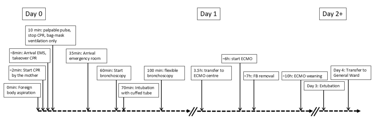
Figure 1Timeline of the case.
DOI: https://doi.org/https://doi.org/10.57187/5263
Diagnosis and treatment of foreign bodies aspirated in the airways is a common challenge, especially in paediatric medicine [1]. While diagnosis may be complicated based on occasionally unspecific clinical presentations, hypoxia-related morbidity and mortality result mostly from critical airway obstruction or complicated bronchoscopic retrieval of a foreign body causing life-threatening respiratory compromise ultimately resulting in respiratory arrest [2].
The ultimate challenges arise when the child presents already out-of-hospital with critical airway obstruction. Under these circumstances, the outcome may depend critically on a well organised chain of experienced pre-clinical and clinical specialists who cooperate in a multidisciplinary and potentially multi-institutional manner.
With this case report, we want to present the management of a 2-year-old child who was about to suffocate following aspiration of a large foreign body into the subglottic space. We describe the “push-down manoeuvre” as a rescue in a “cannot ventilate, cannot oxygenate” situation and the secondary retrieval under extracorporeal membrane oxygenation (ECMO) assistance in a remote centre.
The legal guardians have provided informed consent for the publication of this case report. A girl aged 2 years and 5 months was discovered unresponsive in her room at home, acutely gasping for air. The caregiver observed her choking on an aspirated foreign body and turning blue and unresponsive. Corresponding to the advice provided by the dispatcher of the emergency call centre, the parents initiated cardiopulmonary resuscitation (CPR). On arrival of the emergency medical service, the girl was severely cyanotic, showed severe paradoxical breathing movement and was unconscious. After two minutes of continuous CPR assisted by bag-mask ventilation with 100% oxygen, cyanosis and spontaneous ventilation started to improve. On initial assessment, she presented with a weak pulse and sinus tachycardia. She was urgently rushed to our emergency room. The temporal evolution of events is presented in figure 1.
Figure 1Timeline of the case.
On arrival in the emergency room, she presented clinical signs of severe upper airway obstruction, severe paradoxical breathing movements and hardly any stridor. Her oxygen saturation was in the low nineties while receiving 100% oxygen via a non-rebreather mask. Her capillary blood gas analysis showed profound respiratory acidosis with pH 6.92, pCO2 101 mm Hg, base excess –15.1 mmol/l and bicarbonate 19.8 mmol/l. The lactate level was 8.5 mmol/l. After insertion of peripheral venous access, she was immediately brought to the operating theatre for foreign body retrieval by rigid bronchoscopy as the presentation and the history were highly suggestive of tracheal foreign body aspiration. The parents provided examples of the aspirated foreign body.
Induction of anaesthesia was performed with propofol, remifentanil, atropine, and neuromuscular blockade was initially established using succinylcholine. Bag-mask ventilation via an anaesthesia circle system proved to be difficult. Only small tidal volumes (about 20 ml) could be administered using high inflation pressures of about 30–40 cm H2O.
Tracheal intubation of the child with the rigid bronchoscope proved to be challenging because of difficulties exposing the laryngeal inlet properly under tight time pressure. Rapid and dramatic desaturation occurred immediately after interrupting manual mask ventilation. After the second attempt, we encountered a complete “cannot ventilate-cannot oxygenate” situation and the child’s SpO2 dropped to 10% and heart rate dropped to about 50 bpm. In a desperate attempt after an immediate change of hands of the bronchoscopists, intubation was successful. However, a subglottic foreign body blocked smooth advancement of the bronchoscope into the trachea. The bronchoscopist blindly and forcefully advanced the rigid bronchoscope, attempting to dislodge the subglottic foreign body distally into the mainstem bronchus. Meanwhile, also one dose of adrenaline was administered. Of note, the girl never suffered a cardiac arrest. Thereafter, bag ventilation improved immediately with notably larger tidal volumes (about 120 ml) and oxygenation improved slowly (SpO2 increased to about 70%). Subsequently, the rigid bronchoscope was removed and orotracheal intubation with a cuffed tube was performed without difficulties. Flexible bronchoscopy through the endotracheal tube confirmed that the foreign body had been pushed and wedged into the right mainstem bronchus which was now fully occluded (figure 2).
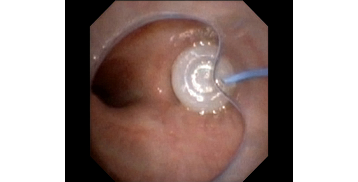
Figure 2Fibreoptic view through the endotracheal tube: foreign body lodged in the right mainstem bronchus after the push-down manoeuvre by the rigid bronchoscope.
Further endoscopic manipulations to extract the foreign body were impossible because of the unstable condition and the lack of sufficient pulmonary reserves. The child had developed frank negative-pressure pulmonary oedema (figure 3).
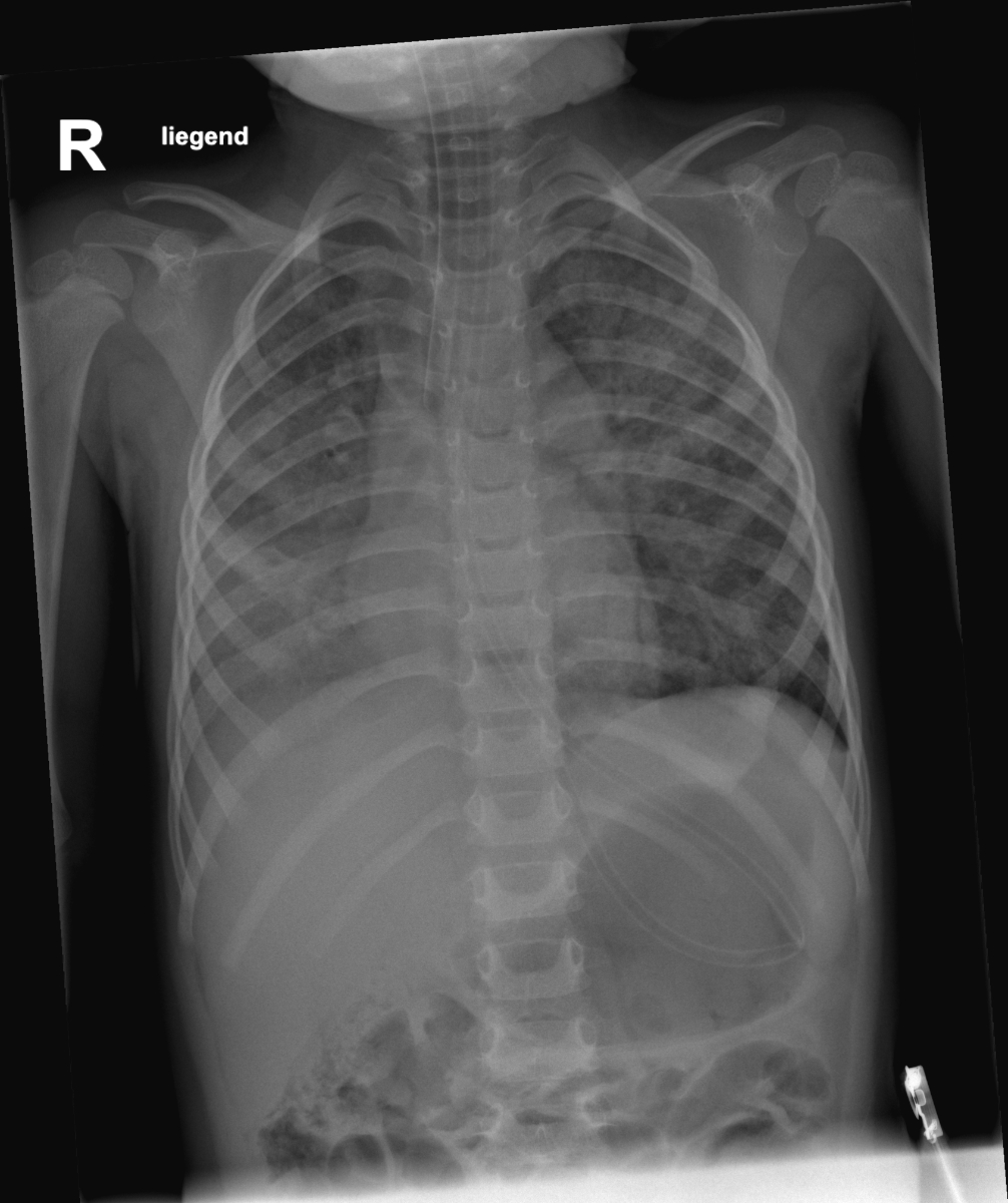
Figure 3Thoracic X-ray after the push-down manoeuvre and intermediate stabilisation with visible negative-pressure pulmonary oedema.
Therefore, air transport to the nearest centre with paediatric ECMO facilities was organised. At the tertiary centre, the patient was placed on veno-venous extracorporeal membrane oxygenation (VV-ECMO) via a 16 Fr bicaval double-lumen catheter due to severe hypoxaemia (PaO₂/FiO₂ ratio of 75). This allowed performing a rigid bronchoscopy safely. It proved difficult to mobilise the foreign body through the vocal cords because of its shape and size (9.8 × 8 × 8 mm) but retrieval was eventually successful. The foreign body was identified as a part of a plastic curtain glider (figure 4).
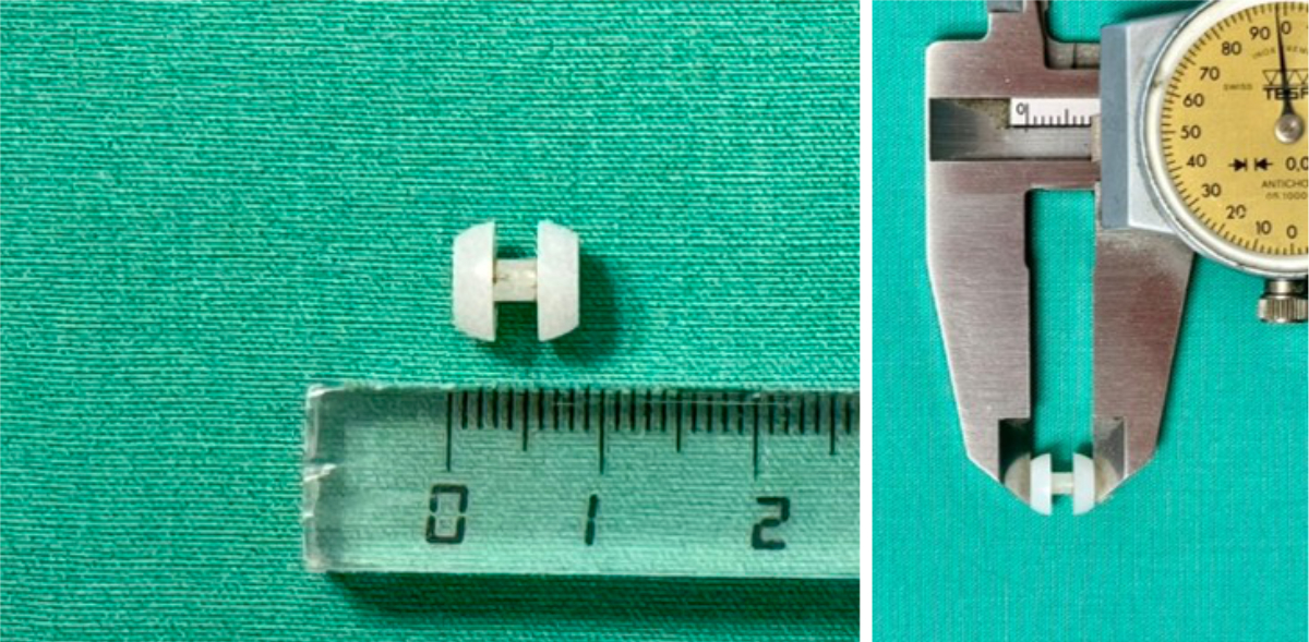
Figure 4Aspirated foreign body (plastic curtain glider).
The patient was easily weaned from ECMO a few hours after foreign body removal and extubated after recovering from the negative-pressure oedema. She was transferred to the regular ward on day four and discharged ten days after the acute aspiration without any neurological sequelae, but with an internal jugular vein thrombosis requiring anticoagulant therapy. One year later, the mother reported normal neurological development of the child.
This case demonstrates the challenges presented by a large foreign body causing near-fatal obstruction of the subglottic area. Foreign body aspiration is a relatively frequent accident and a common reason for presentation to the emergency department. Most aspirated foreign bodies are food particles commonly localised in the right or left mainstem bronchus. Fatal choking events occur most often in children under the age of 5 and are most likely when the foreign body lodges in the larynx or trachea and causes complete airway obstruction [3].
Considering the anaesthetic management in our case, rapid treatment was of the highest importance and rigid bronchoscopy was attempted. We tried to achieve optimal circumstances for removal of the foreign body and therefore started anaesthesia with intravenous propofol and fentanyl with a short trial of positive-pressure ventilation by mask (which was almost impossible) and rapid muscle relaxation. In our case, sevoflurane was not considered an option since ventilation was already minimal at presentation [4].
Bronchoscopy, using either rigid or flexible instruments, is the standard method for foreign body removal from the airway. Rigid bronchoscopy, performed under general anaesthesia or heavy sedation, secures the airway and provides a larger working channel for forceps and other instruments, making it suitable for complex cases. However, critical airway obstruction resulting in hypoxaemia and hypercapnia may lead to rapid deterioration during these procedures. Hence, it is of paramount importance to have alternative plans. In our patient, immediate and safe bronchoscopic removal was not possible due to the size and the location of the foreign body and the occurrence of a “cannot ventilate-cannot oxygenate” situation while preparing for bronchoscopic intubation. The lifesaving manoeuvre was the option to push the foreign body further down into a mainstem bronchus to allow at least one lung ventilation. This technique has been rarely reported but can be of utmost importance since it can lead to cardiopulmonary stabilisation and allow foreign body removal in a second, less dramatic attempt [5].
Unfortunately, our patient also developed negative-pressure pulmonary oedema because of the large negative intrathoracic pressure swings during the forceful inspiratory efforts against the obstruction. The ensuing increase in venous return and left ventricular preload elevates the pressure in the pulmonary veins and causes fluid to leak from the capillaries into the lungs. The pathogenesis of pulmonary oedema associated with upper airway obstruction is multifactorial and beyond the scope of our article. Most cases present within minutes either of the development of acute severe upper airway obstruction or of relief of the obstruction [6]. The severe hypoxaemia prevented any further attempts to retract the foreign body. Therefore, ECMO was considered the best option to stabilise the child before further intervention.
ECMO has been described as an option in life-threatening foreign body aspiration [7, 8]. Two different scenarios are basically described: 1) delayed diagnosis of foreign body aspiration and pulmonary deterioration before, during or after foreign body removal; and 2) rapid respiratory deterioration and need for cardiopulmonary resuscitation with foreign bodies located in the trachea. All ECMO patients described in the literature survived and showed good neurological outcomes – nevertheless we cannot rule out unpublished cases with negative outcomes. Whereas most cases describe patients already situated in a hospital with ECMO capabilities, our case is one of the first to describe a successful transfer to a hospital with ECMO capabilities after near-fatal foreign body aspiration with short-term stabilisation through the “push-down” manoeuvre.
When attempting removal of a critical (sub)glottic or tracheal foreign body, it is of utmost importance to have an experienced anaesthesia and airway team with a backup plan. The push-down manoeuvre can be lifesaving and may allow temporary stabilisation in the event of total airway obstruction. In rare cases of critical pulmonary conditions or technically difficult circumstances for foreign body retrieval, ECMO can secure oxygenation and ventilation and provide enough time for a difficult endoscopic intervention.
Written informed consent was obtained from the legal guardians for the publication of the case report and the accompanying images.
The authors did not receive any financial support for the preparation of this manuscript.
All authors have completed and submitted the International Committee of Medical Journal Editors form for disclosure of potential conflicts of interest. No potential conflict of interest related to the content of this manuscript was disclosed.
1. Saki N, Nikakhlagh S, Rahim F, Abshirini H. Foreign body aspirations in infancy: a 20-year experience. Int J Med Sci. 2009 Oct;6(6):322–8.
2. Huankang Z, Kuanlin X, Xiaolin H, Witt D. Comparison between tracheal foreign body and bronchial foreign body: A review of 1007 cases“, International Journal of Pediatric Otorhinolaryngology, Bd. 76, Nr. 12, S. 1719. Dez. 1725;2012:
3. Chang DT, Abdo K, Bhatt JM, Huoh KC, Pham NS, Ahuja GS. Persistence of choking injuries in children. Int J Pediatr Otorhinolaryngol. 2021 May;144:110685.
4. Black S. „The Anesthetic Considerations of Tracheobronchial Foreign Bodies in Children: A Literature Review of 12,979 Cases. Yearbook of Anesthesiology and Pain Management. 2011 Jan;2011:116–9.
5. Çelenk M, Akyüz Oktay S, Oktay MA. „Lifesaving Maneuver In Full Airway Obstruction Caused By Foreign Body Aspiration: Pushing The Foreign Body To The Right Main Bronchus. Journal of Emergency Medicine Case Reports. 2023 Nov;14(3):71–3.
6. Lang SA, Duncan PG, Shephard DA, Ha HC. Pulmonary oedema associated with airway obstruction. Can J Anaesth. 1990 Mar;37(2):210–8.
7. Anton-Martin P, Bhattarai P, Rycus P, Raman L, Potera R. The Use of Extracorporeal Membrane Oxygenation in Life-Threatening Foreign Body Aspiration: Case Series, Review of Extracorporeal Life Support Organization Registry Data, and Systematic Literature Review. J Emerg Med. 2019 May;56(5):523–9.
8. Ginter D, Johnson KT, Venettacci O, Vanderlaan RD, Gilfoyle E, Mtaweh H. Case report: foreign body aspiration requiring extracorporeal membrane oxygenation. Front Pediatr. 2023 Jul;11:1189722.