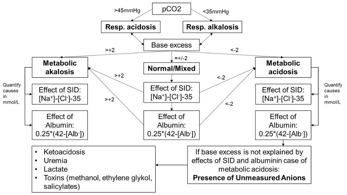
Figure 1Algorithm for the analysis of acid-base status based on Story’s simplified approach.
DOI: https://doi.org/https://doi.org/10.57187/5135
Acid-base disorders are common in acute care settings [1–3]. For example, respiratory acidosis is common in patients with exacerbations of chronic obstructive pulmonary disease while metabolic acidosis is often seen in conditions ranging from sepsis and severe acute or chronic kidney injury to intoxication [4–7]. In most circumstances, the degree of acidosis is a measure of the severity of the underlying disease: for example, lactic acidosis in sepsis is a predictor of adverse outcomes [8]. Under certain conditions, blood gas analysis can be the first indicator of the underlying nature of the patient’s presentation, such as in diabetic ketoacidosis or ethylene glycol poisoning [9, 10]. However, analysis of the metabolic acid-base status can be challenging due to combined disorders resulting in net normal pH or base excess [11]. The physical-chemical approach to the analysis of acid-base disorders, also known as Stewart’s approach, allows a quantitative analysis of the metabolic acid-base status [12]. Story proposed a simplified model of the physical-chemical approach to analyse metabolic acid-base disorders [13].
Acidosis is a common finding in the emergency department with potentially severe underlying causes as outlined above. Nevertheless, there is only little data available on the prevalence, causes and prognosis of severe acidaemia in the emergency department. A recent retrospective analysis focused on emergency patients presenting with a pH <6.9 over a 10-year-period [14]. However, a detailed analysis of the underlying causes of the acid-base disorders was not provided. Another recent retrospective analysis focused on decompensated metabolic acidosis and associated sodium bicarbonate therapy [15]. Again, no details on the underlying causes were presented.
In the following analysis, we aimed to identify the causes of severe acidaemia in the emergency department by structured analysis of the acid-base status, applying the physical-chemical approach as simplified by Story. In addition, we evaluated the outcome of patients with severe acidaemia on admission to the emergency department in terms of hospitalisation, length of hospital stay, ICU admission and in-hospital mortality.
The study was conducted at the Department of Emergency Medicine of Kepler University Hospital in Linz, Austria. Kepler University Hospital Linz is the second largest hospital in Austria with approx. 1850 acute care beds. It is a tertiary care, university institution. The Emergency Department is among the largest in Middle Europe, caring for a median of 272 patients per calendar day in 2024.
The retrospective analysis included all adult patients (i.e. aged ≥18 years) admitted to the Emergency Department with an initial pH <7.15, as measured by a point-of-care acid-base analyser (Radiometer ABL90 Plus), between 1 July 2022 and 30 June 2024.
The following parameters were extracted from the electronic patient record for all identified patients: age, sex, need for intensive care as well as outcome in terms of hospitalisation, length of hospital stay and in-hospital mortality. In addition, blood gas parameters (pH, pCO2, bicarbonate, base excess, anion gap, glucose, sodium, potassium, chloride, calcium) and data from the central laboratory (phosphate, magnesium, albumin, blood urea nitrogen, creatinine, C-reactive protein and haematocrit) were collected. Vital signs, as taken in the Emergency Department, were also used.
For the analysis of patients’ acid-base status in this very specific study, we used the following approach in line with Story’s simplified approach for applying the physical-chemical model. Patients were considered to have respiratory acidosis if pCO2 >45 mm Hg / 6.0 kPa in the initial blood gas analysis, while pCO2 <35 mm Hg / 4.67 kPa was considered respiratory alkalosis. Primary respiratory acidosis was defined as pCO2 >45 mm Hg / 6.0 kPa with concomitant pH <7.35. Primary metabolic acidosis was defined as an acidaemic pH with pCO2 being low to normal (pCO2 <45 mm Hg / 6.0 kPa). Metabolic acid-base status was analysed using Story’s simplified approach: standard base excess was used to quantify the net sum of the components influencing metabolic acid-base status with a value in the range –3 to 3 mmol/l being considered normal. The influence of the electrolytes, expressed as the strong ion difference (SID), was calculated as “[Na+] – [Cl-] - 35” in mmol/l. A value of –2 to 2 mmol/l was considered normal for this analysis. The effect of albumin, the principal weak acid in plasma, was calculated as “0.25 × (42 – [Alb])”. If these factors did not fully explain base excess in patients, the presence of unmeasured anions (UMA) was taken into account. Causes for the presence of unmeasured anions identifiable in this study were ketoacidosis, uraemia and lactic acidosis. Combinations of acid-base disorders, i.e. multiple concurrent acidoses or combinations of acidosis and alkalosis, were considered present if indicated by subcalculations. The algorithm for analysis of the acid-base status was developed by our group on the basis of Story’s simplified approach and is given in figure 1.
Figure 1Algorithm for the analysis of acid-base status based on Story’s simplified approach.
All statistical analyses were performed using JASP version 0.95.0 (JASP Team, University of Amsterdam, The Netherlands). Continuous variables were assessed for distributional characteristics by visual inspection of histograms and Q–Q plots as well as by the Shapiro–Wilk test. Normally distributed continuous variables are presented as mean ± standard deviation (SD), whereas non-normally distributed variables are presented as median with interquartile range (IQR). Categorical variables are reported as absolute counts and percentages.
Given the limited sample size and the exploratory nature of this study, no multivariable regression modelling was performed. Outcome analyses were therefore descriptive and unadjusted. Missing data was handled by complete-case analysis; no imputation was performed. The number of available observations for each variable is reported where relevant.
All statistical tests were two-sided and a p-value <0.05 was considered statistically significant. Due to the exploratory nature of the study, no correction for multiple testing was applied. The study was not powered for hypothesis testing but aimed to provide a comprehensive descriptive and analytical characterisation of severe acidaemia in emergency department patients.
The study was approved by the local ethics committee, the Ethikkommission der Johannes Kepler Universität (Protocol number 1263/2024), and the need for individual patient consent was waived.
The study protocol is available from the study team on demand.
We declare that artificial intelligence was not used in any form in the analysis of data or in the creation of this manuscript.
A total of 63,396 patients presented to the Emergency Department over the study period (1 July 2022 to 30 June 2024), of whom 79 met the inclusion criteria. No patient with a pH <7.15 on venous blood gas analysis was excluded. The median age of patients was 70 years (IQR: 59–82). There was a sex imbalance, with 33 men (58%) and 33 women (42%). Six patients had a positive shock index on admission to the Emergency Department. Seven patients had a mean arterial pressure <65 mm Hg [16].
Blood samples were taken from venous blood. The median pH was 7.08 (IQR: 7.04–7.11) with a minimum of 6.78. The median pCO2 was 41 mm Hg / 5.47 kPa (IQR: 33–78 mm Hg / 4.4–10.4 kPa) with a minimum of 13 mm Hg / 1.73 kPa and a maximum of 164 mm Hg / 21.86 kPa. Median standard bicarbonate was 11 mmol/l (IQR: 8–17 mmol/l) with a minimum of 3.9 mmol/l and a maximum of 30.1 mmol/l. The median standard base excess was -16.3 mmol/l (IQR: -21.85–-5.75) with a minimum of -29.7 mmol/l. Median lactate was 3.6 mmol/l (IQR: 1.8–8.8). Table 1 provides an overview of blood gas analysis data.
Table 1Blood gas parameters of the 79 patients with severe acidaemia on admission to the Emergency Department.
| Parameter | Median (IQR) | Minimum | Maximum |
| pH | 7.08 (7.035–7.11) | 6.78 | 7.15 |
| pCO2 (mm Hg / kPa) | 41/5.48 (24.35–72.85 / 3.25–9.71) | 9.3/1.24 | 145/19.33 |
| Standard bicarbonate (mmol/l) | 11 (8.05–17.1) | 3.9 | 30.1 |
| Sodium (mmol/l) | 137 (134–141) | 115 | 150 |
| Chloride (mmol/l) | 107 (101.5–112) | 77 | 126 |
| Strong ion difference (SID) (mmol/l) | 32 (28–36) | 12 | 43 |
| Anion gap (AG) (mmol/l) | 15.3 (8.95–23.65) | 1.3 | 31.3 |
| Albumin (g/l) | 41 (38.4–47.4) | 15.2 | 53.8 |
| Lactate (mmol/l) | 3.6 (1.8–8.8) | 0.4 | 28 |
| Creatinine (mg/dl) | 1.33 (0.87–3) | 0.24 | 16.5 |
IQR: interquartile range
Approximately half of patients (49%; 39/79) had primary respiratory acidosis, defined as pCO2 >45 mm Hg / 6.0 kPa. Of these, 9 (23%; 11% of all patients) had a normal standard base excess between –3 and 3 mmol/l and only 4 of them had a normal SID of –2 to 2. However, one patient had a combination of lactic acidosis (5.6 mmol/l) and hypoalbuminaemic alkalosis ([Alb] = 15.2 g/l).
Of the 39 patients with respiratory acidosis, 24 (62%) had concomitant metabolic acidosis, expressed by a standard base excess <–3 mmol/l, and 11 had SID-associated acidosis as expressed by “([Na+] – [Cl-] – 35) <–2”. Nearly all (22/24) had lactic acidosis.
Of the 39 patients with respiratory acidosis, 6 (15%) had concomitant metabolic alkalosis, expressed by a standard base excess >3 mmol/l. This was explained by the SID in 3 patients; in the remaining 3, hypoalbuminaemia must be suspected although no measured albumin levels were available.
The remaining half of patients (51%; 40/79) had primary metabolic acidosis, expressed by a normal or low pCO2 (i.e. pCO2 <45 mm Hg / 6.0 kPa). Of these 40 patients, 33 (83%) had concomitant respiratory alkalosis (potentially as a compensation for metabolic acidosis).
Of the 40 patients with primary metabolic acidosis, 23 (58%) had a combination of SID-associated acidosis and UMA acidosis, 8 (20%) had SID acidosis only, 8 (20%) had UMA acidosis only and 1 (2%) had a combination of SID acidosis and metabolic alkalosis caused by hypoalbuminaemia. Figure 2 presents causes of metabolic acidosis on admission to the Emergency Department. The main causes of UMA acidosis were lactic acidosis in 22 patients (44%), diabetic ketoacidosis in 12 (24%) and uraemia in 11 (22%).
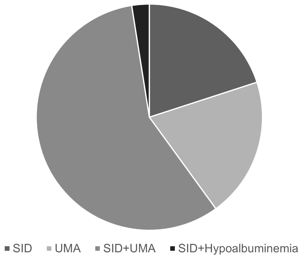
Figure 2Causes of severe metabolic acidosis at presentation to the Emergency Department. SID: strong ion difference; UMA: unmeasured anions.
The cause of UMA acidosis remained unexplained in 2 patients (4%). Figure 3 depicts the causes of UMA acidosis in patients.
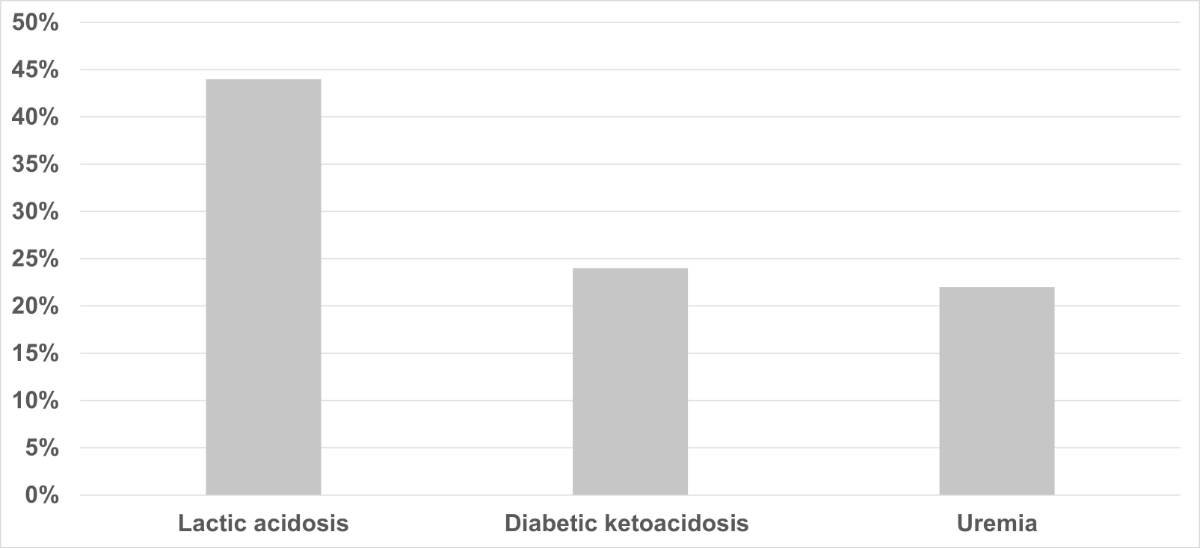
Figure 3Identifiable causes of unmeasured anion acidosis at presentation to the Emergency Department.
One patient with severe respiratory acidaemia was found to have a combined opioid and benzodiazepine intoxication. Acute renal failure was diagnosed in 26 cases. Diabetes mellitus type 1 was diagnosed in 11 patients while diabetes mellitus type 2 was either known or newly diagnosed in 24 patients.
Nearly all patients (96%; 76/79) were hospitalised while three were discharged: the diagnosis was an epileptic seizure in one patient and two patients were discharged against medical advice. The median length of stay of patients was 6 days (IQR: 1–14.5). Thirty patients (38%) were treated in the intensive care unit with a median length of stay of 3.5 days (IQR: 1–9). Eighteen patients (23%) died during hospitalisation, of whom 3 (17%) died in the intensive care unit. In 30 patients (38%), the initial severe acidaemia was not mentioned or discussed in the final medical report.
In the present analysis of severe acidaemia on admission to the Emergency Department, we found that half of the cases were caused by primary respiratory acidosis and the other half by metabolic acidosis. Severe metabolic acidaemia was primarily caused by the effect of strong ion difference or hyperchloraemic acidosis, followed by combined metabolic acidosis with unmeasured anions playing a major role in the development of metabolic acidaemia. Major causes of unmeasured anion acidaemia were lactic acidosis, diabetic ketoacidosis and uraemia. Only one patient had unmeasured anion acidaemia, which could not be further specified retrospectively.
Our study gives a detailed analysis of the causes of severe acidaemia in patients presenting to the Emergency Department. Recently a retrospective analysis from Israel investigated the prognosis of patients with very severe acidaemia with a pH <6.9 [14]. However, there was no detailed analysis of the causes of acidosis in this study. In the present study, only three patients presented with a pH <6.9. Most other studies focused mainly on one specific cause of acidosis or were unspecific. Overall, detailed data on causes and prognosis of severe acidaemia in emergency department patients is scarce and should be a focus of future research.
Strong ion difference-associated metabolic acidosis was quite common in our population. The main cause of this observation was presence of renal insufficiency without uraemia.
By differentiating the causes of acidosis, this study also made another highly important finding: combinations of acid-base disorders are common: 58% of our patients with primary metabolic acidosis had combinations of metabolic acidoses. As was shown in patients with acute liver failure, in whom the acidifying effect of lactic acid was neutralised by the alkalinising effect of hypoalbuminaemia resulting in net normal pH and base excess, a combination of metabolic acid-base disorders can result in concealment of therapeutically relevant acid-base disorders [11]. This is crucial in emergency medicine, where physicians encounter previously unknown and often unresponsive or disorientated patients who may suffer from intoxications, ketoacidosis or uraemia. Blood gas analysis provides an easily accessible and rapid method for diagnosing acid-base disorders. The physical-chemical approach – and especially the easy-to-use method proposed by Story that we adapted into a diagnostic algorithm in the current analysis – might be of great help to the emergency physician in meeting this challenge. Specific teaching sections and standard operating procedures on how to analyse metabolic acid-base disorders could improve reliability and reproducibility of results. This could also result in earlier recognition of severe pathologies such as diabetic ketoacidosis or intoxications. However, further studies are needed to investigate this.
Concerning treatment of patients with severe acidaemia, the administration of sodium bicarbonate is often discussed. A relatively new study published in The Lancet randomised critically ill patients with acidaemia and a pH <7.2 to receive either 4.2% sodium bicarbonate or no sodium bicarbonate [17]; it found no effect on the primary composite outcome of death from any cause by day 28 and the presence of at least one organ failure at day 7. A more recent, larger but retrospective analysis in Australian intensive care units with a pH <7.3 showed a slight mortality reduction with sodium bicarbonate administration [18]. Overall, studies are conflicting with higher quality data indicating no effect of sodium bicarbonate administration, at least in the groups studied [19].
The physical-chemical approach, or Story’s simplified approach as used in the current analysis, represents an alternative model in the analysis of metabolic acid-base status and consequent disorders [13]. In general, the model could be seen as an alternative to rather than an improvement over the traditional model using the anion gap. However, the physical-chemical approach allows for a structured and quantifiable step-by-step analysis of metabolic acid-base disorders.
A 28-year-old woman presented to the emergency department with a history of nausea and non-bloody emesis for two days. She reports not ingesting liquids or solid food for the past 24 hours. The results of the venous blood gas analysis are given in table 2.
Table 2Patient vignette. Results of venous blood gas analysis.
| Parameter | Result |
| pH | 7.164 |
| pCO2 (mm Hg / kPa) | 28/3.7 |
| Standard bicarbonate (mmol/l) | 9.7 |
| Base excess (mmol/l) | –17.9 |
| Sodium (mmol/l) | 137 |
| Chloride (mmol/l) | 107 |
| Albumin (g/l) | 41 |
| Lactate (mmol/l) | 1.3 |
| Creatinine (mg/dl / µmol/l) | 1.6/114 |
Firstly, the impact of electrolytes as quantified by the strong ion difference is calculated:
[Na+] – [Cl-] – 35; i.e. 137 – 107 – 35 = –5
Thus, the strong ion difference explains a total of –5 mmol/l of the base excess of –17.9 mmol/l. Another –12.9 mmol/l remain unexplained.
Secondly, the influence of albumin, a weak acid, needs to be determined:
(42 – [Alb-]) / 4; i.e. (42 – 41) / 4 = 0.25
Consequently, the influence of albumin on base excess is +0.25 mmol/l.
This means that the negative base excess is still unexplained, which indicates metabolic acidosis due to unmeasured anions. In the present case, lactate in the venous blood gas is 1.3 and thus does not explain the remaining –12.9 mmol/l sufficiently. Uraemia appears highly unlikely with a current creatinine level of 1.6 mg/dl / 114 µmol/l and no history of kidney disease. However, ketoacidosis is a possibility given the history of the presenting complaint: severe nausea and vomiting as well as no intake of fluids or solid food. A urine dipstick test was highly positive for the presence of ketone bodies. Therefore, the blood gas analysis of the patient can be classified as metabolic acidaemia caused by (a) ketoacidosis due to starvation (~ –12 mmol/l) and (b) hyperchloraemic acidosis (–5 mmol/l). Of note, the patient’s blood gas analysis normalised after hydration with 5% dextrose.
This study has several limitations that should be considered when interpreting the results. The retrospective, single-centre design inherently limits causal inference and is prone to selection and information bias. Only patients in whom blood gas analysis was performed and documented in the electronic medical record were eligible for inclusion. Consequently, patients with severe acidaemia who did not undergo immediate blood gas analysis may have been missed, potentially leading to an underestimation of the true incidence of severe acidaemia in the emergency department. The sample size was relatively small, reflecting the rarity of severe acidaemia defined by an initial pH <7.15. While sufficient for a detailed descriptive and mechanistic analysis of acid-base disorders, the study was underpowered for robust outcome analyses. All blood gas analyses were performed on venous blood samples. Although venous blood gases are commonly used in emergency medicine and correlate reasonably well with arterial values for pH and bicarbonate, systematic differences exist, particularly for pCO₂. However, it should be noted that the differences with respect to analysis of metabolic acid-base status are clinically negligible. Laboratory data was incomplete in some patients, most notably albumin concentrations. In these cases, hypoalbuminaemic alkalosis was inferred rather than directly quantified, which may have led to misclassification or underestimation of mixed acid-base disorders. Similarly, unmeasured-anion acidosis could not be definitively explained in a small number of patients due to missing or incomplete diagnostic information. No data on urine parameters was analysed in the present study. Finally, the generalisability of the findings may be limited. The study was conducted at a large tertiary care university hospital with a high proportion of critically ill patients, which may not reflect the patient population or case mix of smaller or non-academic emergency departments.
Despite these limitations, the study provides a detailed and clinically relevant analysis of the causes and complexity of severe acidaemia in emergency department patients, highlighting the frequent presence of mixed acid-base disorders and the potential value of a structured physical–chemical approach in acute care settings.
In conclusion, respiratory and metabolic acidoses were found to be equally common as the cause of severe acidosis in patients admitted to the Emergency Department. The most common causes of metabolic acidosis were hyperchloraemic acidosis and unmeasured anions. Lactic acidosis, diabetic ketoacidosis and uraemia were the almost exclusive causes of unmeasured-anion acidosis. Of note, combinations of metabolic acid-base disorders were frequently observed in this group. These findings should raise awareness about not missing therapeutically relevant acid-base disorders in patients admitted to the emergency department.
The data used in this study is available from the authors on justified request.
The authors did not receive any financial support for the preparation of this manuscript.
All authors have completed and submitted the International Committee of Medical Journal Editors form for disclosure of potential conflicts of interest. No potential conflict of interest related to the content of this manuscript was disclosed.
1. Kneidinger N, Funk GC, Lindner G, Drolz A, Schenk P, Fuhrmann V. Unmeasured anions are associated with short-term mortality in patients with hypoxic hepatitis. Wien Klin Wochenschr. 2013 Aug;125(15-16):474–80. doi: https://doi.org/10.1007/s00508-013-0400-9
2. Lindner G, Schwarz C, Grüssing H, Kneidinger N, Fazekas A, Funk GC. Rising serum sodium levels are associated with a concurrent development of metabolic alkalosis in critically ill patients. Intensive Care Med. 2013 Mar;39(3):399–405. doi: https://doi.org/10.1007/s00134-012-2753-3
3. Funk GC, Doberer D, Sterz F, Richling N, Kneidinger N, Lindner G, et al. The strong ion gap and outcome after cardiac arrest in patients treated with therapeutic hypothermia: a retrospective study. Intensive Care Med. 2009 Feb;35(2):232–9. doi: https://doi.org/10.1007/s00134-008-1315-1
4. Lun CT, Tsui MS, Cheng SL, Chan VL, Leung WS, Cheung AP, et al. Differences in baseline factors and survival between normocapnia, compensated respiratory acidosis and decompensated respiratory acidosis in COPD exacerbation: A pilot study. Respirology. 2016 Jan;21(1):128–36. doi: https://doi.org/10.1111/resp.12652
5. Hussain M, Zaki KE, Asef MA, Song H, Treger RM. Unmeasured Organic Anions as Predictors of Clinical Outcomes in Lactic Acidosis due to Sepsis. J Intensive Care Med. 2023 Oct;38(10):975–82. doi: https://doi.org/10.1177/08850666231177602
6. Wang Y, Chen L, Guo G, Gao Y, Gan H. Effect of sodium bicarbonate infusion on hospital mortality in acute kidney injury patients with metabolic acidosis. Front Med (Lausanne). 2023 Oct;10:1268252. doi: https://doi.org/10.3389/fmed.2023.1268252
7. Latus J, Kimmel M, Alscher MD, Braun N. Ethylene glycol poisoning: a rare but life-threatening cause of metabolic acidosis-a single-centre experience. Clin Kidney J. 2012 Apr;5(2):120–3. doi: https://doi.org/10.1093/ckj/sfs009
8. Zhang L, Liu H, Zhang D, Deng Y, Chen X, Zhang L. Non-linear association between lactate and 28 days mortality in elderly patients with sepsis across different SOFA score groups: results from the eICU Collaborative Research Database. Front Med (Lausanne). 2025 Jul;12:1605319. doi: https://doi.org/10.3389/fmed.2025.1605319
9. Umpierrez GE, Davis GM, ElSayed NA, Fadini GP, Galindo RJ, Hirsch IB, et al. Hyperglycemic Crises in Adults With Diabetes: A Consensus Report. Diabetes Care. 2024 Aug;47(8):1257–75. doi: https://doi.org/10.2337/dci24-0032
10. McQuade DJ, Dargan PI, Wood DM. Challenges in the diagnosis of ethylene glycol poisoning. Ann Clin Biochem. 2014 Mar;51(Pt 2):167–78. doi: https://doi.org/10.1177/0004563213506697
11. Funk GC, Doberer D, Fuhrmann V, Holzinger U, Kitzberger R, Kneidinger N, et al. The acidifying effect of lactate is neutralized by the alkalinizing effect of hypoalbuminemia in non-paracetamol-induced acute liver failure. J Hepatol. 2006 Sep;45(3):387–92. doi: https://doi.org/10.1016/j.jhep.2006.03.011
12. Adrogué HJ, Tucker BM, Madias NE. Clinical Approach to Assessing Acid-Base Status: physiological vs Stewart. Adv Chronic Kidney Dis. 2022 Jul;29(4):343–54. doi: https://doi.org/10.1053/j.ackd.2022.04.008
13. Story DA. Stewart Acid-Base: A Simplified Bedside Approach. Anesth Analg. 2016 Aug;123(2):511–5. doi: https://doi.org/10.1213/ANE.0000000000001261
14. Gutgold A, Salameh S, Nashashibi J, Gershinsky Y. Prognosis of patients with extreme acidosis on admission to the emergency department: A retrospective cohort study. Am J Emerg Med. 2024 Feb;76:36–40. doi: https://doi.org/10.1016/j.ajem.2023.10.054
15. Guy C, Holmes NE, Kishore K, Marhoon N, Serpa-Neto A. Decompensated metabolic acidosis in the emergency department: Epidemiology, sodium bicarbonate therapy, and clinical outcomes. Crit Care Resusc. 2023 Jun;25(2):71–7. doi: https://doi.org/10.1016/j.ccrj.2023.05.003
16. Magder SA. The highs and lows of blood pressure: toward meaningful clinical targets in patients with shock. Crit Care Med. 2014 May;42(5):1241–51. doi: https://doi.org/10.1097/CCM.0000000000000324
17. Jaber S, Paugam C, Futier E, Lefrant JY, Lasocki S, Lescot T, et al.; BICAR-ICU Study Group. Sodium bicarbonate therapy for patients with severe metabolic acidaemia in the intensive care unit (BICAR-ICU): a multicentre, open-label, randomised controlled, phase 3 trial. Lancet. 2018 Jul;392(10141):31–40.
18. Blank SP, Blank RM, Laupland KB, Tabah A, Gill D, Kumar A, et al.; Queensland Critical Care Research Network (QCCRN). Sodium bicarbonate administration for metabolic acidosis in the intensive care unit: a target trial emulation. Intensive Care Med. 2025 Jun;51(6):1078–86.
19. Jung B, Jabaudon M, De Jong A, Bitker L, Audard J, Klouche K, et al.; BICARICU-2 Study Group. Sodium Bicarbonate for Severe Metabolic Acidemia and Acute Kidney Injury: The BICARICU-2 Randomized Clinical Trial. JAMA. 2025 Dec;334(22):2000–10. doi: https://doi.org/10.1001/jama.2025.20231