Immunophenotyping of very and
extremely preterm infants at risk of developing sepsis and necrotising
enterocolitis – a prospective, single-centre cohort study
DOI: https://doi.org/https://doi.org/10.57187/4813
Sophie
Strassera,
Valentin von Niederhäuserna,
Noemi Meierb,
Jasmin Simmac,
Claudia Knöpflic,
Emma Slackbde,
Giancarlo Nataluccic,
Johannes Trücka
a Division of
Immunology and Children's Research Center, University Children's Hospital
Zurich, University of Zurich (UZH), Switzerland
b Institute for Food
Sciences, Health and Nutrition, Department of Health Sciences and Technology,
ETH Zurich, Switzerland
c Larsson-Rosenquist Foundation Center for
Neurodevelopment, Growth and Nutrition of the Newborn, Department of
Neonatology, University Hospital Zurich and University of Zurich, Switzerland
d Sir William Dunn
School of Pathology, University of Oxford, Oxford, United Kingdom
e Basel Research Center
for Child Health, Basel, Switzerland
Summary
OBJECTIVE: To characterise adaptive immunity
in very and extremely preterm neonates and assess potential immunological
markers that predispose them to infection.
STUDY DESIGN: Peripheral
blood lymphocytes in 30 very to extremely preterm neonates (mean birth weight
815 g) were analysed using 9-colour flow cytometry at days of life 0–7, 8–14,
and 15–28. Patients were prospectively recruited at the University Hospital of
Zurich from 2021 to 2023. Statistical analyses included group comparisons and
longitudinal assessments.
RESULTS: Six of the thirty preterm neonates
(20%) developed early- or late-onset sepsis, of whom one (3%) also exhibited
signs of necrotising enterocolitis. T and B lymphocytes were
predominantly composed of naïve T cells and unswitched CD27- B cells,
respectively. The CD4/CD8 ratio was initially elevated, especially in extremely
preterm neonates, and declined significantly after week 1. EPT neonates also
had significantly fewer natural killer cells compared with very premature infants.
Although no distinct immunological features were identified as predisposing to
infection, B-cell proportions were significantly higher in infected neonates
during the second week of life, possibly reflecting an immune response to
infection.
CONCLUSION: Adaptive immunity in preterm
neonates is characterised by profound immaturity, including an almost complete
absence of immunological memory and low CD8 T-cell frequency, which may limit
effective pathogen elimination and increase susceptibility to infection. Immunophenotyping
provides valuable insights into the immune processes in this vulnerable
population and, when combined with clinical observations and routine laboratory
findings, could aid in the earlier identification of high-risk neonates and
guide targeted interventions.
Introduction
Preterm infants,
particularly those with very low birth weight and/or gestational age, are highly
susceptible to serious infections such as sepsis and necrotising enterocolitis [1,
2]. This vulnerability is multifactorial, involving
maternal and microbiological factors, but is also linked to the immaturity of
the neonatal immune system [3–7]. Despite a partial understanding of
these factors, detailed insights into the early immune development of very PT
infants, particularly during episodes of suspected or confirmed infection,
remain limited.
Prematurity
and low birth weight are significant risk factors for infection-related
mortality and morbidity in neonates [1, 3, 5], with mortality rates for necrotising
enterocolitis reaching 15–30%, even in high-resource settings [2, 8]. In
Switzerland for example, approximately 0.4%
of children are born with extremely low (<1000 g) birth weight [9], and even higher
rates are reported in other
regions of the world [10]. The immaturity of both innate and adaptive immunity in
these infants
contributes significantly to their vulnerability to infection. At birth, all
newborn infants display deficiencies in T-cell activation, B-cell antibody production,
and T/B-cell interactions [1]. However, PT infants have significantly lower
absolute B- and T-cell counts compared with term infants [3, 4, 11]. Notably, cytotoxic
T cells (CD8+)
are more severely affected than helper T cells (CD4+), leading to an elevated
CD4/CD8 ratio [12–15]. Additionally, T-cells in these infants
are predominantly naïve CD45RA+ T cells, lacking immunological memory [3, 16]. A study
by Scheible et al. showed how
alterations in T-cell maturation in preterm infants might influence respiratory
morbidity in later infancy [17]. B-cell class switching and antibody
production are also impaired in PT infants, partly due to reduced CD40 ligand
expression, which hinders effective T/B-cell interactions [18, 19]. Combined with
lower levels of maternal
immunoglobulin G (IgG) transfer, these immunological features lead to
inefficient phagocytosis and delayed pathogen elimination, elevating the risk of
neonatal infections such as sepsis and necrotising enterocolitis [1, 3, 5].
Necrotising
enterocolitis is characterised by extensive necrosis of intestinal tissue, which
can lead to rupture, bleeding, and subsequent sepsis, and affects 5–10% of
neonates with a birth weight of less than 1500 g [8]. Although the pathogenesis of
necrotising
enterocolitis remains incompletely understood, it likely involves a complex interplay
among microbial colonisation, maternal antibodies, and neonatal immunity [20]. Recent
findings indicate that impaired regulation of intestinal
microbiota – primarily Enterobacteriaceae – by maternal immunoglobulin A
(IgA) leads to gut dysbiosis and is associated with necrotising enterocolitis [21].
Dysregulation in mucosal and peripheral lymphocytes may also play a
role, although evidence for this remains limited [20]. Analysis of resected ileal
tissue from necrotising enterocolitis
patients has revealed altered T-cell differentiation and a reduction in mucosal
regulatory T cells (Tregs), which are crucial for maintaining intestinal
homeostasis, compared with controls [22, 23]. A study by Bochennek et al. further
demonstrated that patients with necrotising
enterocolitis showed reduced peripheral natural killer (NK) cell and naïve
cytotoxic T-cell frequencies [6]. In addition, sepsis in premature infants has been
shown to influence
NK and γδ T-cell frequencies and functional characteristics [24].
A deeper
understanding of peripheral lymphocyte subsets in PT infants, particularly those
with very low birth weight or born extremely preterm, as well as of how
infections such as necrotising enterocolitis and LOS impact these populations,
is needed to enable the early identification of at-risk infants and guide
potential preventive interventions. In this study, multicolour flow cytometry was
used to characterise peripheral lymphocyte populations and their subpopulations
in very preterm infants, both with and without suspected or confirmed sepsis
and/or necrotising enterocolitis.
Methods
Study design and patient
cohort
This is a
substudy of a larger prospective, explorative cohort study conducted at the neonatal
intensive care unit (NICU) department of the University Hospital of Zurich, one
of the major perinatal centres in Switzerland, which provides care for
(extremely) preterm and other high-risk infants. The study was conducted in
collaboration with ETH Zurich from 2021 to 2023. The project involved
collecting biological materials (stool, stomach fluid, urine, and blood) from
very-low-birth-weight infants. The study received approval from the local ethics committee
of Zurich (BASEC
2020-01764) and was performed in accordance with the Declaration of Helsinki. Informed
consent was obtained from the legal guardians of all participating infants. All
neonates with a birth weight <1500 g who were born during the study period and from
whom legal consent for blood
sampling was obtained were eligible. Due to the limited size of this
population, a formal sample size calculation was not feasible. Peripheral blood
samples (100–200μl, EDTA) were
collected serially from 30 very-low-birth-weight infants at different time points
during their stay in
the neonatal intensive care unit (NICU): days of life 1 to 7, DOL 8 to 14, and DOL
15 to 28 (see figure S1 in the appendix). For some patients, additional samples
were also collected from DOL 29 until discharge. Blood sampling was conducted
alongside the collection of other biological materials as part of the
overarching project. For the comparison of immune cell profiles across
different developmental stages, patients were stratified into three gestational
age categories: <26 weeks, 26–28 weeks, and 29–31 weeks.
Sample preparation and
acquisition
Fresh blood
samples were immediately mixed with Cytodelics Whole Blood Stabiliser at the
collection site in a 1:1 ratio, incubated at room temperature for approximately
10 minutes, and subsequently stored at –80°C for later analysis, following the
manufacturer’s protocol (Cytodelics AB; https://www.cytodelics.com/sites/cytodelics.com/files/2023-10/Protocol%20WBCS%20P01%20-%20Sample%20Stabilization_v1.11.pdf) [25, 26]. For staining and analysis, frozen samples
were thawed at 37 °C, then centrifuged in PBS for 4
minutes. Cells were resuspended in 100 μl of a staining mix (adapted Euroflow
panel for main lymphocyte subpopulations) [27–29] and incubated for 30 minutes. Stained
samples were fixed, lysed, and washed according to the Cytodelics Whole Blood
Processing protocol [29]. Data acquisition was performed using a BD
FACSDiva cytometer. Data analysis was conducted with FlowJo software (version
10.10.0) using a gating strategy, as suggested by the Human Immunology Project [30].
Further details on the gating strategy are
provided in figure S2 in the appendix.
Immunophenotyping
The primary
immunodeficiency (PID) orientation tube (Cytognos), a Euroflow primary
immunodeficiency antibody panel, was used with the following
fluorochrome-antibody specificity: CD3 APC; CD4-IgM PerCP-Cy5,5; CD8-IgD FITC;
CD16 PE; CD19-TCRγδ PE-Cy; CD27 BV421; CD45 ACP-C750; CD45RA BV510; CD56 PE.
Additionally, a CD38 BV711 (BD) single antibody was included to enhance B-cell
differentiation. Nine-colour flow cytometry was performed to identify the following
lymphocyte subsets (see also table S1 in the appendix): CD45+ total
lymphocytes; CD45+/CD3+ total T lymphocytes; CD45+/CD3+/TCRγδ-/CD4+ helper T
lymphocytes; CD45+/CD3+/TCRγδ-/CD8+ cytotoxic T lymphocytes; CD45+/CD3+/TCRγδ-/CD4-/CD8-
double-negative T lymphocytes; CD45+/CD19+ total B lymphocytes, and
CD45+/CD16+/CD56+ NK cells. Subpopulations of helper and cytotoxic T cells
included CD27+/CD45RA+ naïve, CD27+/CD45RA- central memory (CM), CD27-/CD45RA-
effector memory (EM), and CD27-/CD45RA+ terminally differentiated (TD) helper T
and cytotoxic T cells, respectively. Within the CD45+/CD19+ B lymphocytes,
CD27- B cells, CD27+/CD38- memory B cells (MBCs), and CD27+/CD38++ plasma cells
(PC) were determined. For CD27- B cells, CD27-/IgM+/IgD+ pre-germinal B cells and
CD27-/IgM-/IgD- switched CD27- B cells were identified. MBCs were further divided into IgM-/IgD- switched MBCs,
IgM++/IgD+ unswitched MBCs, and IgM-/IgD++ MBCs.
Statistical analysis
All statistical
analyses were performed using R (version 2024.09.0+375) with standard packages
for data cleaning, manipulation, and visualisation. Because of the incomplete
availability of absolute lymphocyte counts across
all records (see figure S3 in the appendix for available data and trends in absolute
lymphocyte counts), relative cell frequencies were used for analysis. Data variability
was represented
using medians and interquartile ranges (IQRs). For comparisons between independent
groups, the Mann–Whitney U
test was applied. Paired comparisons, such as changes in cell populations
between visits, were analysed using the Wilcoxon signed-rank test. Associations
between categorical variables, such as gestational age and infection status, were
assessed using Fisher’s exact test. All p-values were two-sided, with
statistical significance defined as p <0.05. Analyses were restricted to
participants with complete data at all three sampling time points. No imputation
was performed for missing values.
Ethics approval
This study
received approval from the local ethics committee of Zurich (BASEC 2020-01764)
and was performed in accordance with the Declaration of Helsinki. Informed consent
was obtained from the legal guardians
of all participating infants.
Results
Patient characteristics
Thirty preterm
neonates were included in this study, with three blood samples collected at three
different time points to represent immunological development in the first month
of life. The cohort consisted of 23 extremely preterm neonates (<26 weeks, n
= 10, 33%; 26–28 weeks, n = 13, 43%) and 7 very preterm neonates (29–31 weeks,
n = 7, 23%). Comprehensive patient data are presented in table 1.
Table 1Patient characteristics. One patient had both
sepsis and suspected necrotising enterocolitis (Bell stage I). Sepsis cases
included four patients with culture-proven sepsis: Staphylococcus epidermidis (n = 2), co-infection with Staphylococcus epidermidis and Staphylococcus caprae (n = 1), and Streptococcus anginosus (n = 1).
Additionally, three patients had negative blood cultures but demonstrated
strong clinical signs of sepsis and received antimicrobial treatment for ≥5
consecutive days. Chorioamnionitis was defined clinically when signs of
intra-amniotic infection were present (e.g. perinatal fever or high infection
parameters of the mother) and/or histologically. Complete antenatal steroids
were defined as ≥2 doses with a 24-hour interval and last dose >24 hours
before birth.
| Features |
All
study participants (n = 30), n (%) |
| Male, n (%) |
12 (40) |
| Gestational age (range), n (%) |
23 weeks (23+3 to 23+5) |
2 (7) |
| 24 weeks (24+3 to 24+5) |
3 (10) |
| 25 weeks (25+0 to 25+6) |
5 (17) |
| 26 weeks (26+2 to 26+4) |
6 (20) |
| 27 weeks (27+0 to 27+6) |
5 (17) |
| 28 weeks (28+4 to 28+5) |
2 (7) |
| ≥29 weeks (29+0 to 31+5) |
7 (23) |
| Birth weight,
g |
Median (range) |
815 (370–1550)
|
| Perinatal
factors, n (%) |
Caesarean
section |
26 (87) |
| Chorioamnionitis
|
15 (50) |
| APGAR at
1’, median (range) |
6 (1–9) |
| APGAR at 10’, median
(range) |
9 (4–10) |
| Complete antenatal
steroids |
20 (66) |
| Endotracheal intubation |
13 (43) |
| Cardiac compression |
1 (3) |
| Adrenaline administration |
0 (0) |
| Infection, n (%) |
Sepsis |
6 (20) |
| Necrotising enterocolitis |
1 (3) |
Main lymphocyte
subpopulations at different gestational ages
For the
analysis of immunological trends during the first month of life (figure 1),
neonates (n = 30) were divided into three gestational age groups: <26 weeks
(n = 10), 26–28 weeks (n = 13), and 29–31 weeks (n = 7). Across all groups, CD3+
T-cell and CD19+ B-cell frequencies remained relatively stable during the first
month. CD56+ NK cells showed significantly lower frequencies at DOL 0–7 in more
preterm infants (< 26 weeks and 26–28 weeks) compared with infants born at ≥29
weeks gestational age (p = 0.049).
However, NK cell counts approached similar frequencies across all age groups at
subsequent time points, with no significant differences noted at later visits. Within
the T-cell population, lower frequencies of CD8+ cytotoxic and higher
frequencies of CD4+ helper T cells were observed at DOL 0–7 in both extremely
preterm groups compared with older neonates. Notably, interindividual variability
within the CD8+ cell group was high. This resulted in an elevated CD4/CD8 ratio
in extremely preterm neonates at DOL ≤7 (median values: <26 weeks = 7.5; 26–28
weeks = 6.9) compared with neonates born ≥29 weeks (median = 3.63). By the
second week of life (DOL 8–14), a significant reduction in the CD4/CD8 ratio was
observed in both extremely preterm groups (<26 weeks: p = 0.027, median = 4.4; 26–28
weeks:
p = 0.017, median = 5.1). No significant
changes of the CD4/CD8 ratio were detected between DOL 8–14 and the final visit
(DOL 15–28). In the B-cell compartment, the majority were CD27- B cells (median
= 98% of total B cells, TBCs), with a small proportion of CD27+/CD38- memory B
cells (median = 1% of TBCs), which predominantly co-expressed IgD/IgM,
indicating an unswitched phenotype. As expected, CD27+/CD38++ plasma cells were
observed at very low frequencies (median = 0.2% of TBCs).
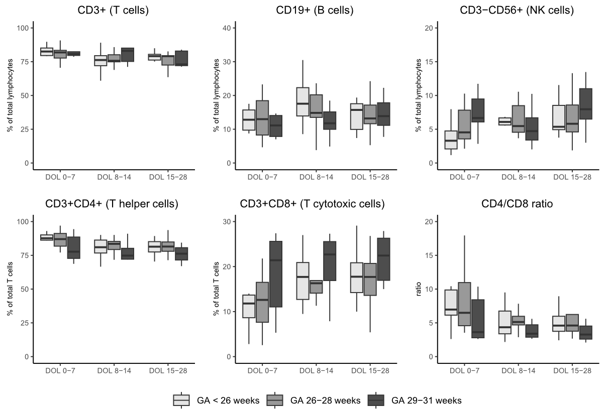
Figure 1Main lymphocyte
populations according to gestational age. Boxes show the interquartile range
(IQR) with the median line; whiskers extend to 1.5×IQR, and outliers beyond
1.5xIQR were excluded for clarity. Abbreviations: DOL, day of life; NK, natural
killer; GA, gestational.
CD4 helper and CD8
cytotoxic cell differentiation in preterm infants
Throughout the
first month of life, the CD4+ and CD8+ T-cell populations in preterm infants were
predominantly composed of naïve T cells (figure 2). This pattern was consistent
across all preterm age groups and time points. The proportion of central memory
T cells in both CD4+ and CD8+ populations ranged between 15% and 20% across all
groups, with no significant temporal changes. Effector memory T cells and terminally
differentiated (TD) T cells within the CD4+ population remained very low throughout
the study period (<1% of CD4+ T cells). By contrast, CD8+ TD T cells exhibited
slightly higher frequencies, with an overall median of 4.7% of CD8+ T cells. One
patient, born at 24+5 weeks of gestational age and without signs of infection,
demonstrated unexpectedly high frequencies of CD8+ TD cells throughout the
first month (DOL 3, 44.9%; DOL 10, 59.5%; DOL 25, 52.0% of CD8+ T cells).
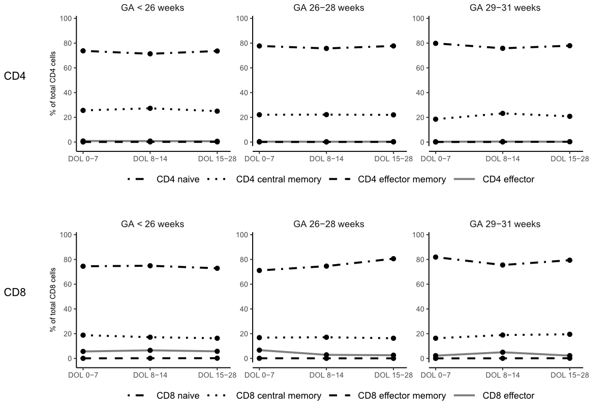
Figure 2Differentiation
of CD4+ and CD8+ T cells across different gestational age groups during the
first month of life. Subpopulations include naïve, central memory (CM),
effector memory (EM), and terminally differentiated (TD) T cells.
Abbreviations: GA, gestational age. Medians were used for data visualisation.
For clarity, variance was not displayed in this figure.
Infections
Six
patients had confirmed or suspected sepsis and/or necrotising enterocolitis
(infection group, see table 1). Among these, four patients had culture-proven
sepsis: Staphylococcus epidermidis
(n = 2; DOL 0 and DOL 11), a co-infection with Staphylococcus epidermidis
and Staphylococcus caprae (n = 1; DOL 17), and Streptococcus anginosus (n = 1; DOL 0). The patient with Staphylococcus caprae sepsis also exhibited signs of necrotising
enterocolitis (Bell stage 1) at DOL 11. Two patients were treated with
antibiotics for ≥5 consecutive days without pathogen identification and were therefore
classified as having culture-negative sepsis. One patient with clinical
suspicion of sepsis at DOL 69, who was treated with antibiotics for <5 days
without pathogen identification, was excluded from the infection group. No
significant correlations were found between gestational age or birth weight
groups and infection risk. Longitudinal data on lymphocyte subpopulations are shown
in figure 3. Notably, one patient with sepsis onset at birth (DOL 0) displayed
the highest B-cell frequency (53% of all lymphocytes) at DOL 5, possibly in
response to infection, with a gradual normalisation over the first 3 weeks of
life (DOL 10, 30%; DOL 18, 15%). Although no definitive trends or correlations
were observed with infection onset, the infection group had significantly
higher B-cell frequencies at DOL 8–14 compared with neonates without infection
(p = 0.04, medians: infection group 20%; healthy group 14%). Both groups
exhibited a gradual increase in CD8+ T cells and a corresponding decrease in
the CD4/CD8 ratio during the first month of life. However, this trend reached
statistical significance only in the healthy group between DOL 0–7 and DOL 8–14.
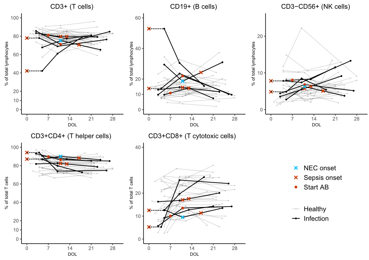
Figure 3Main lymphocyte
subsets in preterm neonates with and without infection. Main lymphocyte subsets
are shown for preterm neonates with sepsis and/or necrotising enterocolitis
(infection group) compared with those without infection. Infection onset is
indicated to provide context for observed immunological changes. For the two
patients with culture-negative sepsis, the exact onset of infection was
unclear; therefore, the initiation of antibiotic therapy (lasting ≥5 consecutive
days) is marked instead. Abbreviations: DOL, day of life; NK, natural killer;
NEC, necrotising enterocolitis; AB, antibiotics
Discussion
In our explorative
cohort of thirty very preterm neonates with birthweight ≤1550 g, flow cytometric
analysis of peripheral lymphocyte populations in the first month of life
revealed a considerable immaturity of adaptive immunity, with naïve T cells
(both CD4+ and CD8+) and unswitched CD27- B cells dominating the T- and B-cell
compartment, respectively. No distinct immunological features predisposing infants
to infection were observed; however, infants with infections exhibited
significantly higher B-cell proportions in the second week of life, possibly indicating
an immunological response to previous or ongoing infection.
Consistent
with findings from previous studies [3, 4], the CD4+ and CD8+ T-cell
populations in preterm neonates were almost completely devoid of immunological
memory, with extremely low proportions of EM and TD T cells. This deficiency may
contribute to their vulnerability to infection, as efficient pathogen
recognition and elimination remain limited. However, term neonates have also been
shown to express similarly high naïve T-cell proportions (albeit in higher
absolute numbers) [31–33], suggesting that the immaturity of
CD4+ and CD8+ T cells may be due to limited postnatal antigenic stimulation
rather than a feature unique to preterm birth. Notably, lower gestational age
was associated with reduced CD8+ T-cell frequencies during the first week of
life, resulting in an increased CD4/CD8 ratio, particularly in extremely
preterm neonates [11–15]. In this context, Bochennek et al. demonstrated a
significant correlation between low naïve CD8+ T-cell frequencies and necrotising
enterocolitis occurrence [6], but this effect was not observed in our study,
and its clinical significance remains unclear.
Within the B-cell
compartment, we observed extremely low frequencies of memory and plasma B cells,
in line with limited immunoglobulin (Ig) production in preterm neonates, making
these infants highly dependent on passively transferred maternal Ig. However, IgG
transfer is reduced in preterm neonates, as it is interrupted by preterm delivery
[34], thereby impairing IgG-mediated pathogen
recognition and elimination. Another study showed that IgA-mediated control of
intestinal microbiota often fails in preterm and low-birth-weight infants, contributing
to the pathogenesis of necrotising enterocolitis [17]. Interestingly, preterm
neonates with sepsis and/or necrotising enterocolitis had significantly higher B-cell
proportions in their second week of life, likely as a humoral immune response
to a previous or ongoing infection. However, further longitudinal studies are
needed to clarify this finding in a larger sepsis cohort.
Additionally,
we assessed NK cells and found extremely preterm neonates expressed
significantly fewer NK cells in their first week of life compared to older neonates.
Although extremely preterm infants were generally at a higher risk for
infection, no statistically significant correlation was observed between low NK
cell frequencies and infection risk in our study. In contrast, Bochennek et al.
found that low NK cell frequencies significantly increased the risk of late-onset
sepsis [6], suggesting that NK cell-mediated mechanisms of innate immunity may
play a role in neonatal susceptibility to infection.
Several
limitations should be considered in this study. First, the sample sizes were
relatively small, limiting statistical power. Second, clinical features such as
chorioamnionitis, severe respiratory distress/intubation, or mode of delivery
may have influenced immunological processes but could not be statistically
addressed due to low sample sizes. Third, for logistical reasons, samples were stabilised
with a whole blood stabiliser and then frozen for weeks to months before data
acquisition, which may have reduced sample quality, although the stabiliser kit
has been validated in previous studies [26, 27]. However, obtaining blood
samples from preterm neonates, particularly those with extremely low birth weights,
is challenging, and few longitudinal data on lymphocyte phenotyping in this
population exist. Last, as whole-blood samples were not routinely collected alongside
study samples, few data on absolute lymphocyte counts exist, and the analysis
was performed using frequencies rather than absolute numbers. Even though the
use of relative frequencies has drawbacks (e.g. limited information on small
populations and difficult-to-interpret changes in populations separately), it
provides important insights into immune cell composition and dynamics and how
these influence individual susceptibility to infection. Despite these limitations,
this study is among the few to assess peripheral blood samples rather than cord
blood or resected tissue in this difficult-to-study population.
In
conclusion, the immunophenotyping of very and extremely preterm neonates during
their first month of life highlighted how immaturity within both T- and B-cell
subsets contributes to their heightened susceptibility to infection. Although no
distinct immunological profiles predictive of infection were identified,
certain features associated with extreme preterm birth, such as a high CD4/CD8
ratio and low NK cell frequency, may contribute to the multifactorial
pathogenesis of neonatal infections. These findings warrant further studies to identify
high-risk immunological constellations in this extremely vulnerable population.
Data availability statement
Due to the
sensitive nature of patient data and ethical considerations, the underlying
dataset cannot be publicly shared. Deidentified aggregated data, along with
data dictionaries, where applicable, and the analytical code are available from
the corresponding author upon reasonable request for scientific purposes
consistent with the approved ethical framework.
Acknowledgments
We thank all on-site nurses and other medical staff
for their support throughout this study. Their contributions were essential and
greatly appreciated.
Author contributions: Study
concept and design: VvN, JT, ES, NM, SS; acquisition and analysis of data: JS,
CK, NM, SS, VvN, JT; interpretation of data: SS, JT, ES, GN; drafting of
manuscript: SS, JT; revision of manuscript: SS, VvN, ES, GN, JT
Johannes
Trück, MD DPhil
Division of Immunology and Children's Research Center
University Children's Hospital Zurich
Lenggstrasse 30
CH-8008 Zürich
johannes.trueck[at]kispi.uzh.ch
References
1. Melville JM, Moss TJ. The immune consequences of preterm birth. Front Neurosci. 2013 May;7:79.
10.3389/fnins.2013.00079
2. Fitzgibbons SC, Ching Y, Yu D, Carpenter J, Kenny M, Weldon C, et al. Mortality of
necrotizing enterocolitis expressed by birth weight categories. J Pediatr Surg. 2009 Jun;44(6):1072–5.
10.1016/j.jpedsurg.2009.02.013
3. Huenecke S, Fryns E, Wittekindt B, Buxmann H, Königs C, Quaiser A, et al. Percentiles
of Lymphocyte Subsets in Preterm Infants According to Gestational Age Compared to
Children and Adolescents. Scand J Immunol. 2016 Nov;84(5):291–8. 10.1111/sji.12474
4. Walker JC, Smolders MA, Gemen EF, Antonius TA, Leuvenink J, de Vries E. Development
of lymphocyte subpopulations in preterm infants. Scand J Immunol. 2011 Jan;73(1):53–8.
10.1111/j.1365-3083.2010.02473.x
5. Collins A, Weitkamp JH, Wynn JL. Why are preterm newborns at increased risk of infection? Arch
Dis Child Fetal Neonatal Ed. 2018 Jul;103(4):F391–4. 10.1136/archdischild-2017-313595
6. Bochennek K, Fryns E, Wittekindt B, Buxmann H, Quaiser A, Fischer D, et al. Immune
cell subsets at birth may help to predict risk of late-onset sepsis and necrotizing
enterocolitis in preterm infants. Early Hum Dev. 2016 Feb;93:9–16. 10.1016/j.earlhumdev.2015.10.018
7. Wynn JL, Wong HR. Pathophysiology and treatment of septic shock in neonates. Clin
Perinatol. 2010 Jun;37(2):439–79. 10.1016/j.clp.2010.04.002
8. Battersby C, Santhalingam T, Costeloe K, Modi N. Incidence of neonatal necrotising
enterocolitis in high-income countries: a systematic review. Arch Dis Child Fetal
Neonatal Ed. 2018 Mar;103(2):F182–9. 10.1136/archdischild-2017-313880
9 Bundesamt für Statistik (BFS). Säuglingssterblichkeit und Gesundheit der Neugeborenen
im Jahr 2022. BFS Aktuell 2023;14 Gesundh:1–8.
10 Organization WH. Born too soon: the global action report on preterm birth n.d.
11. Berrington JE, Barge D, Fenton AC, Cant AJ, Spickett GP. Lymphocyte subsets in term
and significantly preterm UK infants in the first year of life analysed by single
platform flow cytometry. Clin Exp Immunol. 2005 May;140(2):289–92. 10.1111/j.1365-2249.2005.02767.x
12. Qazi KR, Bach Jensen G, van der Heiden M, Björkander S, Holmlund U, Haileselassie Y,
et al. Extremely Preterm Infants Have Significant Alterations in Their Conventional
T Cell Compartment during the First Weeks of Life. J Immunol. 2020 Jan;204(1):68–77.
10.4049/jimmunol.1900941
13. Sampah ME, Hackam DJ. Dysregulated Mucosal Immunity and Associated Pathogeneses in
Preterm Neonates. Front Immunol. 2020 May;11:899. 10.3389/fimmu.2020.00899
14. Govindaraj D, Jensen GB, Rahman Qazi K, Sverremark-Ekström E, Abrahamsson T, Jenmalm MC.
Effects of extremely preterm birth on cytokine and chemokine responses induced by
T-cell activation during infancy. Clin Transl Immunology. 2024 May;13(5):e1510. 10.1002/cti2.1510
15. Kamdar S, Hutchinson R, Laing A, Stacey F, Ansbro K, Millar MR, et al. Perinatal inflammation
influences but does not arrest rapid immune development in preterm babies. Nat Commun.
2020 Mar;11(1):1284. 10.1038/s41467-020-14923-8
16. Juretić E, Juretić A, Uzarević B, Petrovečki M. Alterations in lymphocyte phenotype
of infected preterm newborns. Biol Neonate. 2001;80(3):223–7. 10.1159/000047147
17. Scheible KM, Emo J, Laniewski N, Baran AM, Peterson DR, Holden-Wiltse J, et al. T
cell developmental arrest in former premature infants increases risk of respiratory
morbidity later in infancy. JCI Insight. 2018 Feb;3(4):1–17. 10.1172/jci.insight.96724
18. Nonoyama S, Penix LA, Edwards CP, Lewis DB, Ito S, Aruffo A, et al. Diminished expression
of CD40 ligand by activated neonatal T cells. J Clin Invest. 1995 Jan;95(1):66–75.
10.1172/jci117677 doi: https://doi.org/10.1172/JCI117677
19. Kaur K, Chowdhury S, Greenspan NS, Schreiber JR. Decreased expression of tumor necrosis
factor family receptors involved in humoral immune responses in preterm neonates.
Blood. 2007 Oct;110(8):2948–54. 10.1182/blood-2007-01-069245
20. Denning TL, Bhatia AM, Kane AF, Patel RM, Denning PW. Pathogenesis of NEC: role of
the innate and adaptive immune response. Semin Perinatol. 2017 Feb;41(1):15–28. 10.1053/j.semperi.2016.09.014
21. Gopalakrishna KP, Macadangdang BR, Rogers MB, Tometich JT, Firek BA, Baker R, et al. Maternal
IgA protects against the development of necrotizing enterocolitis in preterm infants.
Nat Med. 2019 Jul;25(7):1110–5. 10.1038/s41591-019-0480-9
22. Weitkamp JH, Koyama T, Rock MT, Correa H, Goettel JA, Matta P, et al. Necrotising
enterocolitis is characterised by disrupted immune regulation and diminished mucosal
regulatory (FOXP3)/effector (CD4, CD8) T cell ratios. Gut. 2013 Jan;62(1):73–82. 10.1136/gutjnl-2011-301551
23. Lu P, Struijs MC, Mei J, Witte-Bouma J, Korteland-van Male AM, de Bruijn AC, et al. Endoplasmic
reticulum stress, unfolded protein response and altered T cell differentiation in
necrotizing enterocolitis. PLoS One. 2013 Oct;8(10):e78491. 10.1371/journal.pone.0078491
24. Rahman Qazi K, Jensen GB, van der Heiden M, Björkander S, Marchini G, Jenmalm MC,
et al. Extreme prematurity and sepsis strongly influence frequencies and functional
characteristics of circulating γδ T and natural killer cells. Clin Transl Immunology.
2021 Jun;10(6):e1294. 10.1002/cti2.1294
25. Olin A, Henckel E, Chen Y, Lakshmikanth T, Pou C, Mikes J, et al. Stereotypic Immune
System Development in Newborn Children. Cell. 2018 Aug;174(5):1277–1292.e14. 10.1016/j.cell.2018.06.045
26. Carter MJ, Fish M, Jennings A, Doores KJ, Wellman P, Seow J, et al. Peripheral immunophenotypes
in children with multisystem inflammatory syndrome associated with SARS-CoV-2 infection.
Nat Med. 2020 Nov;26(11):1701–7. 10.1038/s41591-020-1054-6
27. van der Velden VH, Flores-Montero J, Perez-Andres M, Martin-Ayuso M, Crespo O, Blanco E,
et al. Optimization and testing of dried antibody tube: the EuroFlow LST and PIDOT
tubes as examples. J Immunol Methods. 2019 Dec;475:112287. 10.1016/j.jim.2017.03.011
28. van der Burg M, Kalina T, Perez-Andres M, Vlkova M, Lopez-Granados E, Blanco E, et
al. The EuroFlow PID orientation tube for flow cytometric diagnostic screening of
primary immunodeficiencies of the lymphoid system. Front Immunol. 2019 Mar;10:246.
10.3389/fimmu.2019.00246
29. Blanco E, Pérez-Andrés M, Arriba-Méndez S, Contreras-Sanfeliciano T, Criado I, Pelak O,
et al.; EuroFlow PID group. Age-associated distribution of normal B-cell and plasma
cell subsets in peripheral blood. J Allergy Clin Immunol. 2018 Jun;141(6):2208–2219.e16.
10.1016/j.jaci.2018.02.017
30. Maecker HT, McCoy JP, Nussenblatt R. Standardizing immunophenotyping for the Human
Immunology Project. Nat Rev Immunol. 2012 Feb;12(3):191–200. 10.1038/nri3158
31. Melville JM, Moss TJ. The immune consequences of preterm birth. Front Neurosci. 2013 May;7:79.
10.3389/fnins.2013.00079
32. Walker JC, Smolders MA, Gemen EF, Antonius TA, Leuvenink J, de Vries E. Development
of lymphocyte subpopulations in preterm infants. Scand J Immunol. 2011 Jan;73(1):53–8.
10.1111/j.1365-3083.2010.02473.x
33. van Duuren IC, van Hengel ORJ, Penders J, Duijts L, Smits HH, Tramper-Stranders GA.
The developing immune system in preterm born infants: From contributor to potential
solution for respiratory tract infections and wheezing. Allergy: European Journal
of Allergy and Clinical Immunology 2024. https://doi.org/10.1111/all.16342.
34. Wessel RE, Dolatshahi S. Quantitative mechanistic model reveals key determinants of
placental IgG transfer and informs prenatal immunization strategies. PLOS Comput Biol.
2023 Nov;19(11):e1011109. 10.1371/journal.pcbi.1011109
Appendix
The appendix is available in the pdf version of the article at https://doi.org/10.57187/4813.