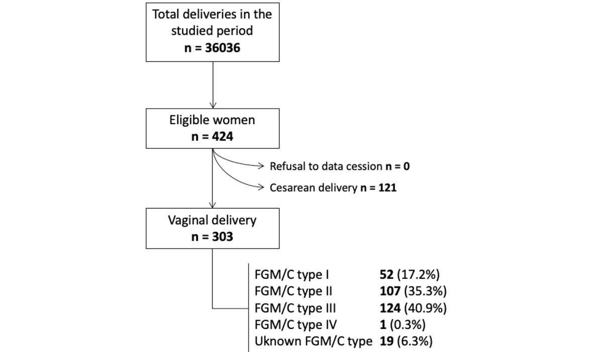
Figure 1 Flowchart of eligible participants.
DOI: https://doi.org/https://doi.org/10.57187/4785
The World Health Organization (WHO) defines female genital mutilation/cutting (FGM/C) as the partial or total removal of the female external genitalia as well as any genital lesion with no medical indication [1], although this definition has been debated by many authors [2]. Official statistics from 2022 estimated that 84,650,032 women had undergone female genital mutilation/cutting [3]. UNICEF recently estimated that approximately 230 million women and girls are living with female genital mutilation/cutting worldwide [4]. In Switzerland, indirect estimates suggest that approximately 24,600 women and girls are living with or at risk of female genital mutilation/cutting [5].
Female genital mutilation/cutting can lead to various psychophysical consequences, including genitourinary, psychological, sexual, and obstetric issues. Regarding the latter, it may increase the risk of episiotomy, perineal tears, instrumental or caesarean deliveries, postpartum haemorrhage, prolonged labour, and obstetric anal sphincter injuries [6–11]. Pschophysical comorbidities are exacerbated by discrimination and suboptimal care due to a lack of training on the management of female genital mutilation/cutting, among other factors [12].
Defibulation after female genital mutilation/cutting type III (infibulation, narrowing of the vaginal orifice by apposition of the labia, with or without excision of the clitoris) can reduce obstetrical risks [6]. Women who have undergone defibulation, possibly along with mid-lateral episiotomy, experience fewer spontaneous lacerations, postpartum haemorrhages, and third- and fourth-degree lacerations [9].
Our study aimed primarily to develop a predictive model of the risk of obstetric anal sphincter injuries in women with female genital mutilation/cutting delivering vaginally. In addition, we sought to provide a comprehensive descriptive analysis of this population and their obstetric outcomes.
A retrospective observational study was conducted at the Maternity Hospital in Geneva University Hospitals (HUG), a Swiss tertiary university hospital with over 4000 annual births and a specialised outpatient clinic for women and girls with female genital mutilation/cutting established in 2010.
The study aimed to develop a predictive model for obstetric anal sphincter injuries (main outcome) in women with female genital mutilation/cutting who underwent vaginal delivery. Secondary objectives included evaluating the predictive capacity of specific variables for this outcome, with a focus on the mode of delivery and the presence of defibulation. The obstetric population of women with female genital mutilation/cutting at the centre was also described.
Women with female genital mutilation/cutting who were admitted for delivery to the Maternity Ward at HUG from 1 April 2010 (coinciding with the inauguration of the specialised female genital mutilation/cutting clinic and the initiation of professional training for healthcare providers) to 31 December 2018 were included. Exclusion criteria were written refusal of consent for data use in research or contraindications to vaginal delivery unrelated to female genital mutilation/cutting.
Medical records of eligible women were reviewed to collect data on demographics, medical and obstetric history, pregnancy outcomes, and labour and delivery details, as well as data specifically on perineal tears and postpartum complications. Ethical approval was secured from the cantonal ethics committee prior to data collection (Commission Cantonale d'Éthique de la Recherche ID 2019-00999), which waived the need for informed consent.
Data were anonymised and entered into the Redcap® database. Female genital mutilation/cutting types were categorised according to the WHO international classification (table 1), and obstetric injuries affecting the anal sphincter were considered “obstetric anal sphincter injuries” (i.e. third- and fourth-degree perineal lacerations). Candidate predictors were also pre-specified based on existing literature and clinical plausibility and included maternal age, parity, body mass index, gestational age at delivery, history of perineal tear, mode of delivery (spontaneous, instrumental, or caesarean), foetal weight estimation, timing of defibulation, episiotomy, and use of anaesthesia. Missing data are reported in the tables and were not imputed.
To develop a model with 10 predictors for a 10% prevalence of severe perineal tears, a minimum sample size of 380 women was calculated. However, due to the study’s retrospective design and electronic record accessibility, all eligible women were included in the analysis.
Table 1WHO classification of female genital mutilation/cutting.
| WHO classification of female genital mutilation/cutting |
| Type I - Partial or total removal of the prepuce (Ia), with possible cutting of the glans of the clitoris (Ib). |
| Type II - Excision: Partial or total removal of the inner labia (IIa), with possible cutting of the glans of the clitoris (IIb) and the outer labia (IIc). |
| Type III - Infibulation: Narrowing of the vaginal opening through the creation of a covering seal by apposition of the inner labia (IIIa) or outer labia (IIIb), with or without cutting of the clitoris. |
| Type IV - All other harmful procedures: Pricking, piercing, incising, scraping, and cauterising the genital area. |
The occurrence of a severe perineal tear led to the formation of two sub-cohorts. Continuous variables were described using medians and interquartile ranges, and categorical variables were described using proportions. Group comparisons were made using the Wilcoxon and Fisher’s exact tests.
A predictive model was attempted using stepwise logistic regression, with p-values of 0.05 for inclusion and 0.10 for exclusion of variables. The final model’s predictive ability was evaluated using the area under the receiver operating characteristic (ROC) curve.
The HUG prenatal clinic screens all pregnant women from countries with prevalent female genital mutilation/cutting practices. If a woman reports female genital mutilation/cutting, or if it is detected during medical history intake or a vulvar examination, she is referred to the specialised clinic, where she receives detailed information, undergoes a specialised examination, and discusses and records her birth plan. Preventive information is also provided if a female child is expected. In cases of female genital mutilation/cutting type III, defibulation options are discussed as early as possible: the patient can choose whether she prefers to undergo the procedure during the 2nd trimester (under local or spinal anaesthesia) or during delivery, while she is in the first stage of labour. Additionally, she has the choice to undergo complete (up to the clitoris) or partial (above the urethra) defibulation. Preoperative discussions cover vulvar anatomy and expected changes post-defibulation, such as quicker urination. Partners can join these consultations if desired and if the woman consents. We respectfully decline requests for reinfibulation [13, 14], offering information and counselling instead, and allowing the woman to choose between a defibulation uncovering the urethra and vagina only, or also the clitoris once she is informed of the possibilities and potential advantages. Certified trained female interpreters are available for non-French- or English-speaking patients, who represent 30% of our patients. Information and counselling on defibulation include drawings, videos, 3D models, and a web app, which explain the surgery, associated care, pain control, and changes in anatomy and physiology [15–17].
Since the clinic's inception, several improvements have been made in diagnosing, recording, and coding female genital mutilation/cutting and managing complications. Our electronic obstetric medical records now include a dedicated section for female genital mutilation/cutting history and descriptions [18]. In addition, regular training on female genital mutilation/cutting and defibulation is provided to residents and midwives through simulation [13]. An agreed written delivery plan is established for each patient; episiotomy is not routine. If a patient has chosen defibulation during labour, the on-call gynaecologist performs it during the first stage. For caesarean deliveries, those with non-defibulated female genital mutilation/cutting type III can opt to have defibulation concurrently.
During the postpartum period, trained midwives provide pain management and care, particularly for episiotomies, tears, or recent defibulation, including care to prevent adhesions. A follow-up appointment at the female genital mutilation/cutting clinic 6 to 8 weeks after delivery is offered to assess healing, provide additional information and support, and discuss childbirth, prevention, and general postpartum care.
From April 2010 to December 2018, a total of 36,036 women delivered at the Maternity Hospital at HUG, of whom 424 (1.18%) had female genital mutilation/cutting (40.9% were type III female genital mutilation/cutting). Of these, 303 (71.5%) had a vaginal birth and 121 (28.5%) had a caesarean delivery. Among the 303 women who had a vaginal delivery, 174 (57.4%) experienced a perineal tear, of whom 12 (4.0%) had an obstetric anal sphincter injury. Fifteen (6.5%) patients had a history of obstetric anal sphincter injury in a prior delivery, but no recurrence of obstetric anal sphincter injury was found in our group. The episiotomy rate was 29.1% and was higher in women with obstetric anal sphincter injuries (58%) than in those without them (28%). Instrumentation occurred in 46 (15.2%) of vaginal deliveries, resulting in 4 cases of obstetric anal sphincter injuries (8.7%). The relative risk of obstetric anal sphincter injury associated with non-spontaneous delivery was 2.79 (95% CI 0.88-8.90; p = 0.091). The characteristics of the included participants, overall and according to the type of perineal lesion, are described in figure 1 and table 2.
Table 2Characteristics of the participants, according to the type of perineal lesion at delivery.
| Variable | All participants | No OASI | OASI | p-value |
| n = 303 | n = 291 (94.04%) | n = 12 (9.96%) | ||
| Maternal age – years | 29.59 (8.32) | 29.60 (8.26) | 27.36 (10.64) | 0.5781 |
| Nulliparous | 78 (25.74%) | 71 (24.40%) | 7 (58.33%) | 0.0152 |
| Height – cm | 163 (7) | 163 (7) | 160 (7) | 0.0321 |
| … Missing values | 1 (0.3%) | |||
| Weight at delivery – kg | 73 (17) | 73 (17) | 65.5 (11.5) | 0.0231 |
| … Missing values | 22 (7.3%) | |||
| BMI at delivery – kg/m2 | 27.36 (6.07) | 27.45 (5.94) | 24.84 (4.64) | 0.1261 |
| … Missing values | 22 (7.3%) | |||
| Gestational age at delivery – days | 280 (12) | 280 (12) | 281 (10) | 0.3511 |
| Hypertensive disorders | 12 (4.00%) | 12 (4.17%) | 0 (0%) | >0.9992 |
| … Missing values | 3 (1.0%) | |||
| Diabetes mellitus | 31 (10.33%) | 31 (10.76%) | 0 (0%) | 0.6212 |
| … Missing values | 3 (1.0%) | |||
| Cephalic presentation | 297 (99.66%) | 285 (99.65%) | 12 (100%) | >0.9992 |
| … Missing values | 5 (1.7%) | |||
| Spontaneous onset of labour | 186 (62.00%) | 178 (61.81%) | 8 (66.67%) | >0.9992 |
| … Missing values | 3 (1.0%) | |||
| Locoregional anaesthesia | 202 (67.33%) | 192 (66.67%) | 10 (83.33%) | 0.3492 |
| … Missing values | 3 (1.0%) | |||
| Desinfibulation | 104 (34.90%) | 101 (35.07%) | 3 (30%) | >0.9992 |
| … Antenatal | 77 (25.84%) | 75 (74.26%) | 2 (66.67%) | >0.9992 |
| … Intrapartum | 27 (9.06%) | 26 (25.74%) | 1 (33.33%) | |
| … Missing values | 5 (1.7%) | |||
| Episiotomy | 88 (29.14%) | 81 (27.93%) | 7 (58.33%) | 0.0452 |
| … Missing values | 1 (0.3%) | |||
| Postpartum haemorrhage (≥ 500 ml) | 24 (7.97%) | 23 (7.96%) | 1 (8.33%) | >0.9992 |
| … Missing values | 2 (0.7%) | |||
| Neonatal weight – g | 3345 (525) | 3335 (515) | 3420 (580) | 0.7061 |
| … Missing values | 1 (0.3%) | |||
| Apgar score at 5th minute | 10 (0) | 10 (0) | 10 (1) | 0.3501 |
| … Missing values | 4 (1.3%) | |||
| Umbilical artery pH | 7.23 (0.08) | 7.23 (0.08) | 7.19 (0.06) | 0.1081 |
| … Missing values | 42 (14.2%) | |||
| Admission to NICU | 8 (2.66%) | 8 (2.77%) | 0 (0%) | >0.9992 |
| … Missing values | 2 (0.7%) | |||
OASI: obstetric anal sphincter injury; BMI: body mass index; NICU: neonatal intensive care unit.
Continuous variables are presented as the median (interquartile range). Categorical variables are presented as n (%).
1 Wilcoxon rank-sum test
2 Fisher’s exact test
Figure 1 Flowchart of eligible participants.
When excluding women with a prior vaginal delivery, the prevalence of obstetric anal sphincter injuries was 8.4% (7/83). No differences were observed in the analysed variables of this subgroup between women who did and did not have an obstetric anal sphincter injury. A table with complete information is available in the Appendix (table S1).
In our population of women with female genital mutilation/cutting type III, 73.1% of the patients were defibulated prior to delivery, either before (47.3%) or during the current pregnancy (25.7%). For the latter, defibulation was performed as an intrapartum procedure in 85.2% of vaginal deliveries (23 women) and in 87.5% of caesarean deliveries (14 women). Six women with female genital mutilation/cutting type III who had a vaginal delivery experienced a severe tear (4.8%), among whom three (50.0%) had undergone defibulation (one having undergone an intrapartum procedure during the current pregnancy). There were no differences in the risk of obstetric anal sphincter injuries according to the timing of defibulation (p >0.99).
Because of the low prevalence of severe perineal tears within our cohort, it was not possible to develop a predictive model, as there were not enough events to identify adequate predictors. Among the 12 women affected by obstetric anal sphincter injuries, a higher proportion were nulliparous (58.3% in the obstetric anal sphincter injuries group vs 24.4% in the group without; p = 0.015) and had undergone an episiotomy (7 women, 58.3%, vs 81 women, 27.9%, who did not experience a sphincter injury; p = 0.045). A stratified analysis considering parity and the performance of an episiotomy revealed that nulliparous women were at an increased risk of severe perineal tears. However, episiotomy neither increased nor reduced the risk of obstetric anal sphincter injury in either group when classified by parity (figure 2). Foetal weight did not differ significantly between groups.
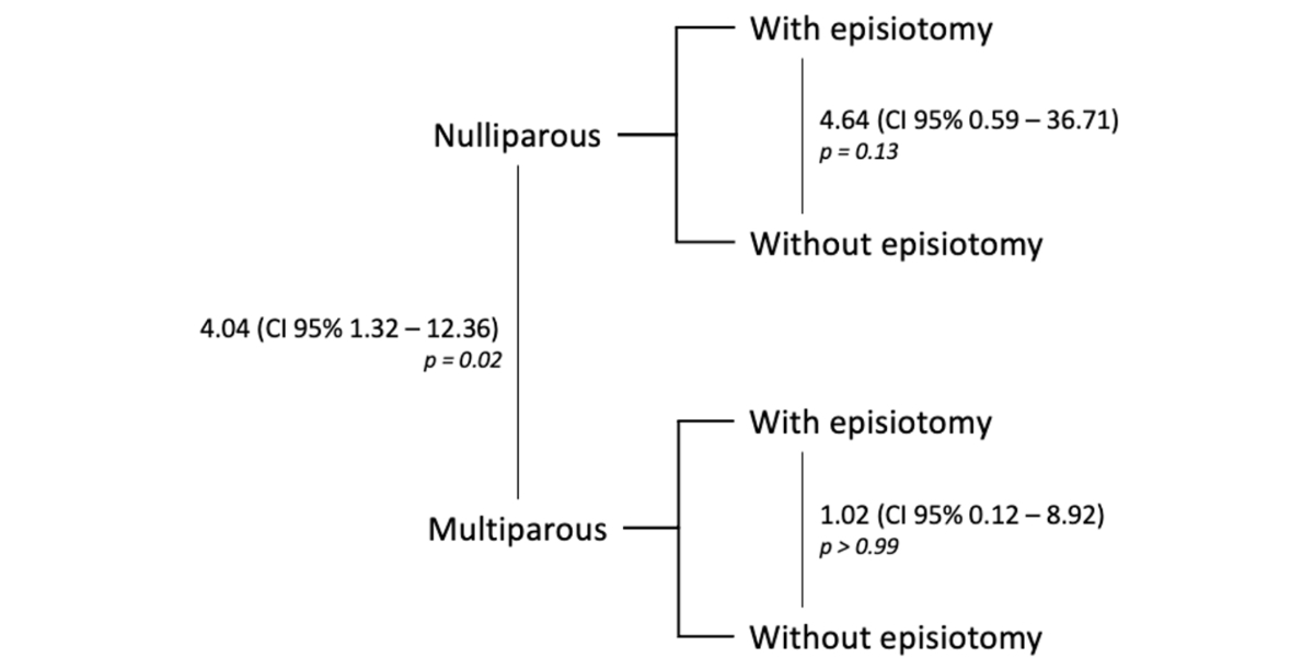
Figure 2 Relative risks of obstetric anal sphincter injury based on parity and the performance of an episiotomy; p-values were computed using Fisher’s exact test.
Of the women who experienced an obstetric anal sphincter injury, seven (58.3%) had female genital mutilation/cutting type III. Three of them had undergone defibulation (one in labour, one before pregnancy, and one during a previous delivery). For the remaining four women, information regarding defibulation was not available.
The overall caesarean delivery rate was 28.54% (121/424 participants), of which 25.62% (31/121) were elective procedures. The median maternal age in these women was higher than that among women undergoing vaginal delivery (32.22 vs. 29.59 years; p <0.001). Twelve (11.54%) women had a history of perineal tear, among whom two had a previous obstetric anal sphincter injury. The prevalence of female genital mutilation/cutting type III in women with caesarean birth was 49.6% (compared with 40.9% in vaginal deliveries; p = 0.315). The results comparing the modes of delivery are shown in the Supplementary Materials (table S2 in the appendix).
When analysing the indication for caesarean delivery, none were primarily performed due to female genital mutilation/cutting or related conditions, although the most common cause for caesarean delivery was a previous caesarean section (iterative indication). Other primary indications included arrest of labour at any stage or suspected foetal distress. A complete list of indications for caesarean births is available in the Supplementary Materials (Table S3).
Our study was initially designed as a prediction model study to estimate the risk of obstetric anal sphincter injury among women with female genital mutilation/cutting, with the ultimate goal of supporting clinical decision-making during labour. This objective was motivated by previous reports showing a relatively high prevalence of obstetric anal sphincter injury (around 7.4%) in similar populations [19], which may lead caregivers to avoid vaginal deliveries or to approach them with excessive caution, often resulting in more medicalised births.
However, in our cohort, the observed prevalence of obstetric anal sphincter injury was 4%, a rate comparable to that of the general obstetric population (3.4%) [20, 21]. The unexpectedly low number of events meant that the development of a robust prediction model was not statistically feasible. Accordingly, our analyses focused on descriptive outcomes, which nonetheless provide clinically relevant insights into the obstetric course of women with female genital mutilation/cutting in a specialised care setting.
Within these descriptive analyses, we identified parity and the performance of an episiotomy as potential contributing factors to obstetric anal sphincter injury. Nulliparity is a well-established determinant of obstetric anal sphincter injury in women without female genital mutilation/cutting [20], but large-scale population studies have reported conflicting results in women with female genital mutilation/cutting [22, 23]. The role of episiotomy also remains debated, with evidence suggesting both protective [22] and neutral [21] effects. In our cohort, episiotomy was more frequent among nulliparous women; however, stratified analyses indicated that episiotomy did not reduce the risk of obstetric anal sphincter injury in either nulliparous or multiparous women. These findings are consistent with those in the general population, in which nulliparity increases obstetric anal sphincter injury risk, but episiotomy does not confer protection [24, 25] and may even increase the risk in multiparous women [20].
We did not find an association between the timing of defibulation and the occurrence of an obstetric anal sphincter injury. In the case of female genital mutilation/cutting type III, defibulation is necessary to allow vaginal delivery and it is not associated with increased obstetrical adverse events [11]. In our cohort, both antenatal and intrapartum defibulation appeared safe and were not associated with an increased risk of perineal injuries. These findings are consistent with WHO recommendations [26], which emphasise that women should be informed about both options and empowered to choose according to their preferences. From our clinical experience, antenatal defibulation is often preferred to allow healing and adaptation before delivery, whereas intrapartum defibulation may be chosen to avoid additional pregnancy procedures and to benefit from obstetric anaesthesia [27].
The caesarean delivery rate in our cohort (28.5%) was similar to the regional rate (31.6% – data from the Swiss Federal Statistics Office). As expected, rates were higher among women without prior vaginal deliveries, consistent with previous meta-analyses [6]. Importantly, none of the caesareans in our study were indicated solely because of female genital mutilation/cutting. Even if we acknowledge that professionals may be biased by the genital lesion when deciding on the best mode of delivery, all caesareans had a primary medical cause.
The primary limitation of our study is the insufficient number of events to fully address its main objective. Although this low incidence is reassuring clinically, it limited our ability to provide a practical predictive tool for clinicians in settings without specialised care. Additionally, the retrospective design of the study, relying on data from clinical records, introduced the potential for missing information, which hinders bias control and may result in misinterpretation of the findings. Furthermore, the study lacks a comparison group of women without female genital mutilation/cutting, and no pre-implementation data were available to evaluate changes following the introduction of the structured care model in 2010. However, the study was designed to describe outcomes within a specific population receiving standardised care, making internal validity and clinical relevance within this group the primary focus.
The main strength of our study is the relatively large cohort of women with female genital mutilation/cutting followed at a single centre with standardised protocols and trained staff. This consistency reduces retrospective bias and reflects the effectiveness of a specialised care model in ensuring safe obstetric outcomes for this population.
Our institution has extensive and long-standing experience in the follow-up of women with female genital mutilation/cutting during pregnancy. Our specialised clinic is led by a gynaecologist who assesses the type of female genital mutilation/cutting, explains the findings to the patient and, eventually, to family members, discusses the birth plan, and establishes primary, secondary, and tertiary prevention strategies to improve patients’ quality of life. If a language barrier is present, we can involve a certified female interpreter during consultations to provide clear explanations, which helps reduce stress and avoid complications in the delivery room [28]. Additionally, all staff involved in prenatal care and in the labour ward, including midwives, residents, and senior obstetricians, undergo regular training in recognising and managing female genital mutilation/cutting. They are also trained in how to conduct labour and assist deliveries in this specific context.
Although we were unable to develop a predictive model, our findings suggest that with appropriate identification, antenatal counselling, defibulation when indicated, and care provided by trained professionals, obstetric outcomes among women with female genital mutilation/cutting can be comparable to those in the general population [29]. Future prospective studies are warranted to further explore not only obstetric risks but also broader outcomes such as sexual function, mental health, and quality of life.
Although our study did not yield a prediction model due to the low number of obstetric anal sphincter injuries, it highlights that, within a structured model of care, women with female genital mutilation/cutting can experience favourable perinatal outcomes. These results underscore the importance of individualised, respectful, and evidence-based management during pregnancy and childbirth.
Data can be shared upon reasonable request to the corresponding author.
Author contributions: FM, LF, and JA developed the original idea, wrote the research protocol, and planned the study. LF collected the data. FM oversaw the analysis, which was also discussed by LF, JA, and BMT. FM and LF wrote the manuscript, which was corrected and approved by JA and BMT. FM: Conceptualisation, formal analysis, methodology, investigation, validation, writing – original draft, writing – review and editing. LF: Conceptualisation, data curation, investigation, validation, writing – review and editing. BMT: Investigation, supervision, validation, writing – review and editing. JA: Conceptualisation, investigation, supervision, validation, writing – review and editing.
This research did not receive any specific grant from funding agencies in the public, commercial, or not-for-profit sectors.
All authors have completed and submitted the International Committee of Medical Journal Editors form for disclosure of potential conflicts of interest. No potential conflict of interest related to the content of this manuscript was disclosed.
1. Soins pour les filles et les femmes vivant avec des mutilations sexuelles féminines: manuel clinique [Care of women and girls living with female genital mutilation: a clinical handbook]. Genève: Organisation mondiale de la Santé; 2020.
2. United Nations Children’s Fund. Female Genital Mutilation/Cutting: A global concern. New York: UNICEF; 2016.
3. Cottler-Casanova S, Horowicz M, Gayet-Ageron A, Abdulcadir J. Female genital mutilation/cutting (FGM/C) coding capacities in Swiss university hospitals using the International Classification of Diseases (ICD). BMC Public Health. 2021 Jun;21(1):1151.
4. UNICEF. Female genital mutilation (FGM) [Internet]. WHO; 2024 [cited 2024 Apr 19]. https://data.unicef.org/topic/child-protection/female-genital-mutilation/. Accessed 19 Apr 2024
5. Swiss Federal Office of Public Health. Measures against female genital mutilation [Internet]. 2023 [cited 2024 Apr 19]. https://www.bag.admin.ch/bag/en/home/strategie-und-politik/nationale-gesundheitsstrategien/gesundheitliche-chancengleichheit/chancengleichheit-in-der-gesundheitsversorgung/massnahmen-gegen-weibliche-genitalverstuemmelung.html. Accessed 19 Apr 2024
6. Sylla F, Moreau C, Andro A. A systematic review and meta-analysis of the consequences of female genital mutilation on maternal and perinatal health outcomes in European and African countries. BMJ Glob Health. 2020 Dec;5(12):e003307.
7. Lurie JM, Weidman A, Huynh S, Delgado D, Easthausen I, Kaur G. Painful gynecologic and obstetric complications of female genital mutilation/cutting: A systematic review and meta-analysis. PLoS Med. 2020 Mar;17(3):e1003088.
8. Berg RC, Underland V. Obstetric Consequences of Female Genital Mutilation/Cutting (FGM/C) [Internet]. Oslo, Norway: Knowledge Centre for the Health Services at The Norwegian Institute of Public Health (NIPH); 2013 [cited 2024 Apr 19]. http://www.ncbi.nlm.nih.gov/books/NBK464778/. Accessed 19 Apr 2024
9. Balachandran AA, Duvalla S, Sultan AH, Thakar R. Are obstetric outcomes affected by female genital mutilation? Int Urogynecol J. 2018 Mar;29(3):339–44.
10. Davis G, Jellins J. Female genital mutilation: obstetric outcomes in metropolitan Sydney. Aust N Z J Obstet Gynaecol. 2019 Apr;59(2):312–6.
11. Bonavina G, Spinillo SL, Sotiriadis A, Bulfoni A, Kaltoud R, Salvatore S, et al. Effect of type III female genital mutilation on obstetric outcomes: A systematic review and meta-analysis. Heliyon. 2024 Apr;10(8):e29336.
12. Johnson-Agbakwu CE, Michlig GJ, Koukoui S, Akinsulure-Smith AM, Jacobson DS. Health outcomes and female genital mutilation/cutting: how much is due to the cutting itself? Int J Impot Res. 2023 May;35(3):218–27.
13. Abdulcadir J, Marras S, Catania L, Abdulcadir O, Petignat P. Defibulation: A Visual Reference and Learning Tool. J Sex Med. 2018 Apr;15(4):601–11.
14. Abdulcadir J, McLaren S, Boulvain M, Irion O. Health education and clinical care of immigrant women with female genital mutilation/cutting who request postpartum reinfibulation. Int J Gynaecol Obstet. 2016 Oct;135(1):69–72.
15. WebApp Let’s Talk FGM/C - Sciences. Sexes, Identités - UNIGE [Internet]. 2021 [cited 2024 Apr 19]. https://www.unige.ch/ssi/tuiles/webapp-lets-talk-mgf/. Accessed 19 Apr 2024
16. Abdulcadir J, Dewaele R, Firmenich N, Remuinan J, Petignat P, Botsikas D, et al. In Vivo Imaging-Based 3-Dimensional Pelvic Prototype Models to Improve Education Regarding Sexual Anatomy and Physiology. J Sex Med. 2020 Sep;17(9):1590–602.
17. Desinfibulation - ENGLISH [Internet]. 2021 [cited 2024 Apr 19]. https://www.youtube.com/watch?v=S9UWBmDBzac. Accessed 19 Apr 2024
18. Horowicz M, Cottler-Casanova S, Abdulcadir J. Diagnoses and procedures of inpatients with female genital mutilation/cutting in Swiss University Hospitals: a cross-sectional study. Reprod Health. 2022 May;19(1):104.
19. Wuest S, Raio L, Wyssmueller D, Mueller MD, Stadlmayr W, Surbek DV, et al. Effects of female genital mutilation on birth outcomes in Switzerland. BJOG. 2009 Aug;116(9):1204–9.
20. Klokk R, Bakken KS, Markestad T, Holten-Andersen MN. Modifiable and non-modifiable risk factors for obstetric anal sphincter injury in a Norwegian Region: a case-control study. BMC Pregnancy Childbirth. 2022 Apr;22(1):277.
21. Eshraghi B, Hermansson J, Berggren V, Marions L. Risk of obstetric anal sphincter tear among primiparous women with a history of female genital mutilation, giving birth in Sweden. PLoS One. 2022 Dec;17(12):e0279295.
22. Rodriguez MI, Seuc A, Say L, Hindin MJ. Episiotomy and obstetric outcomes among women living with type 3 female genital mutilation: a secondary analysis. Reprod Health. 2016 Oct;13(1):131.
23. Female genital mutilation and obstetric outcome: WHO collaborative prospective study in six African countries. The Lancet. Elsevier; 2006;367:1835–41. https://doi.org/
24. Mahgoub S, Piant H, Gaudineau A, Lefebvre F, Langer B, Koch A. Risk factors for obstetric anal sphincter injuries (OASIS) and the role of episiotomy: A retrospective series of 496 cases. J Gynecol Obstet Hum Reprod. 2019 Oct;48(8):657–62.
25. Jiang H, Qian X, Carroli G, Garner P. Selective versus routine use of episiotomy for vaginal birth - Jiang, H - 2017 | Cochrane Library. [cited 2024 Sept 18]; https://www.cochranelibrary.com/cdsr/doi/10.1002/14651858.CD000081.pub3/full?contentLanguage=en. Accessed 18 Sept 2024
26. World Health Organization. WHO guidelines on the management of health complications from female genital mutilation [Internet]. Geneva: World Health Organization; 2016 [cited 2024 Apr 20]. https://iris.who.int/handle/10665/206437. Accessed 20 Apr 2024
27. Taraldsen S, Vangen S, Øian P, Sørbye IK. Risk of obstetric anal sphincter injury associated with female genital mutilation/cutting and timing of deinfibulation. Acta Obstet Gynecol Scand. 2022 Oct;101(10):1163–73.
28. Schrot-Sanyan S, Kolanska K, Haimeur Y, Varlas V, Parisot-Liance L, Daraï E, et al. Language barrier as a risk factor for obstetric anal sphincter injury - A case-control study. J Gynecol Obstet Hum Reprod. 2021 Oct;50(8):102138.
29. Cinelli H, Lelong N, Lesclingand M, Alexander S, Blondel B, Le Ray C, et al. Female genital mutilation/cutting in women delivering in France: An observational national study. Int J Gynaecol Obstet Off Organ Int Fed Gynaecol Obstet; 2024.
The appendix is available in the pdf version of the article at https://doi.org/10.57187/4785.