Toxoplasmosis in focus – how a small parasite
conquers the world
DOI: https://doi.org/https://doi.org/10.57187/4379
a Spital
Thurgau, Münsterlingen, Switzerland
Summary
Toxoplasma
gondii is one of the
most important parasites globally in both veterinary and human medicine, with
significant impacts on livestock. Approximately one-third of the global population
is latently infected. In humans, infection is typically asymptomatic or causes mononucleosis-like
symptoms, but primary infection during pregnancy and reactivation in
immunocompromised individuals are of particular concern. Latent infection may be associated
with personality and neuropsychiatric
disorders or neurodegenerative diseases. Despite a sound rationale, many congenital
toxoplasmosis surveillance
programmes have been discontinued due to limited effectiveness. Current One Health
research focuses on developing new therapies and
vaccines to prevent transmission in humans and animals.
Introduction
Toxoplasmosis is among the most common zoonoses
worldwide. The infection is caused by the protozoan parasite Toxoplasma
gondii, one of the most successful parasites globally. A significant
portion of the world’s population, as well as many animals, is latently
infected without being aware of it, but most people become infected later in life
through contaminated food.
The initial infection is almost always
asymptomatic. In rare cases, it may present with flu-like symptoms, including
fever, peripheral lymphadenopathy, and prolonged fatigue. Thereafter, the
pathogen remains dormant as a tissue cyst within the host. Of particular
concern is primary infection during pregnancy in humans and in animals, which
can lead to miscarriage or foetal damage. Reactivation of latent toxoplasmosis
can be extremely dangerous for immunocompromised individuals. Moreover, T.
gondii has been implicated in behavioural changes in infected animals and
may play a significant role in the development of neurological and psychiatric
disorders in humans.
Global patterns and transmission pathways of
Toxoplasma gondii
History and seroprevalence
T. gondii was first identified in 1907 by the French
physicians and parasitologists Charles Nicolle and Louis H. Manceaux. Its name
derives from the Greek words «toxon» (bow, arc) and «plasma»
(formation, shape), as well as from the North African rodent gundi, in
which the parasite was first discovered. It was not until 1948 that the
parasite was identified in humans [1, 2].
Humans, like other warm-blooded animals, act as
intermediate hosts for T. gondii worldwide. Members of the family Felidae
(domestic and wild cats) are the definitive hosts. The expansion of agriculture
over the past 11,000 years, along with the establishment of the cat–mouse
transmission cycle, likely facilitated the global spread of T. gondii and the selection of specific
parasite lineages. [3]. Although only a limited number of T. gondii
genotypes predominate in the Northern Hemisphere, a wide variety of genotypes coexist
in South America. Moreover, South American strains tend to exhibit higher
virulence than those found in the Northern Hemisphere [3].
The parasite is one of the most important foodborne
zoonoses globally and a major cause of production losses in agriculture [1, 2]. Depending
on the region, 5–70% of humans are
latently infected, alongside livestock and rodents [1, 2, 4, 5]. Human seroprevalence
is estimated to be around 32% in Europe, 15% in North America, 45% in
South America, 54% in Australia, 42% in Africa, and 25% in Asia. Within a
single country, prevalence can vary widely across demographic groups depending
on culture, socioeconomic status, and diet [4, 5].
Over recent decades, T. gondii
seroprevalence has declined in industrialised countries, likely due to reduced
environmental exposure [4, 5]. Among women of childbearing age, rates dropped
from 53% to 35% in Switzerland (1982–1999), from 84% to 23% in France
(1960–2006), and from 43.3% to 31.5% in Austria (1995–2012) [4–6]. Similar
declines have occurred in indoor-raised livestock, while prevalence has increased
in free-range animals [1, 2, 10]. Despite improved diagnostics, global data
remain fragmented, with substantial variation by region, time, and methodology.
In many countries, surveillance is limited, and prevalence data are outdated or
incomplete [4].
Pathogenesis and environmental resistance
Toxoplasma gondii belongs to the phylum Apicomplexa,
which is closely related to Plasmodium and Cryptosporidium. Apicomplexa are single-celled eukaryotic parasites with a complex life cycle involving
sexual reproduction in definitive hosts and asexual reproduction in
intermediate hosts. They exploit host cells for efficient propagation [1, 2].
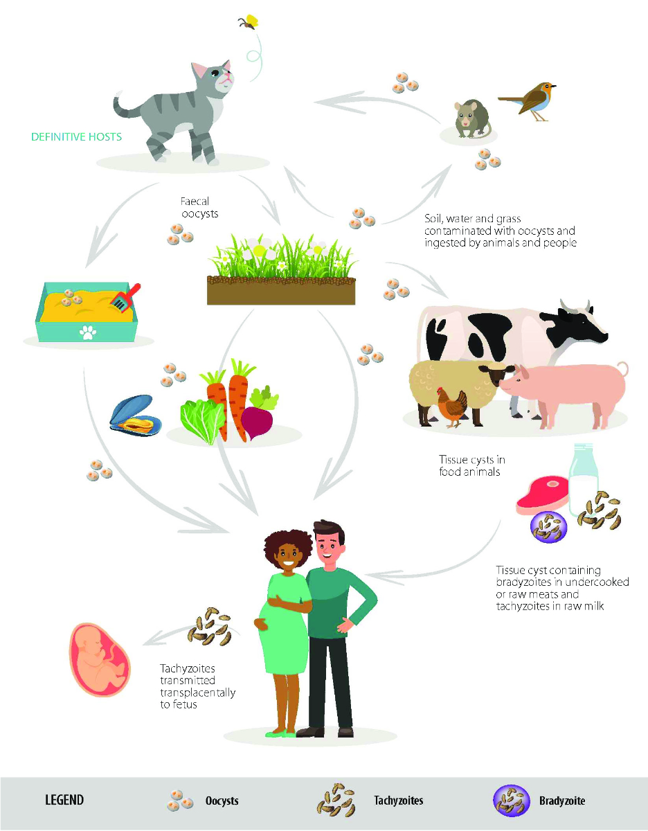
Figure 1
Foodborne transmission pathways for Toxoplasma gondii. From: EFSA Panel on Biological Hazards (BIOHAZ); Koutsoumanis K, Allende A, Alvarez-Ordóñez
A, Bolton D, Bover-Cid S, Chemaly M, et al. Public health risks associated with food-borne
parasites. EFSA J. 2018 Dec 4;16(12):e05495. doi: 10.2903/j.efsa.2018.5495, published
under the license CC BY-ND 4.0.
Cats, the definitive hosts, shed vast numbers of
oocysts in their faeces (3 to 810 million), which contaminate soil, plants, and
water [8]. In warm, moist environments, the process of sporulation is initiated,
and infectious sporozoites develop within the oocysts [9]. Sporozoites are resistant
to heat and desiccation and can survive for months in the environment [10].
Intermediate hosts ingest these sporulated oocysts through contaminated food or
prey. Humans can acquire the parasite through contaminated water, unpasteurised
milk, vegetables, and undercooked meat, or through contact transmission during gardening,
cleaning cat litter, or playing
in sandboxes [10] (figure 1). Transmission via human breast milk has not been demonstrated
(though tachyzoites are excreted in the milk of infected livestock), and direct
human-to-human transmission is not possible, except through organ transplants
or blood transfusions [1].
In the stomach and intestines, the oocysts
dissolve, releasing tachyzoites that invade host cells. A characteristic apical
complex, the conoid, aids in cell invasion (figure 2). Current research, including
research conducted in Switzerland, focuses on understanding this mechanism [7].
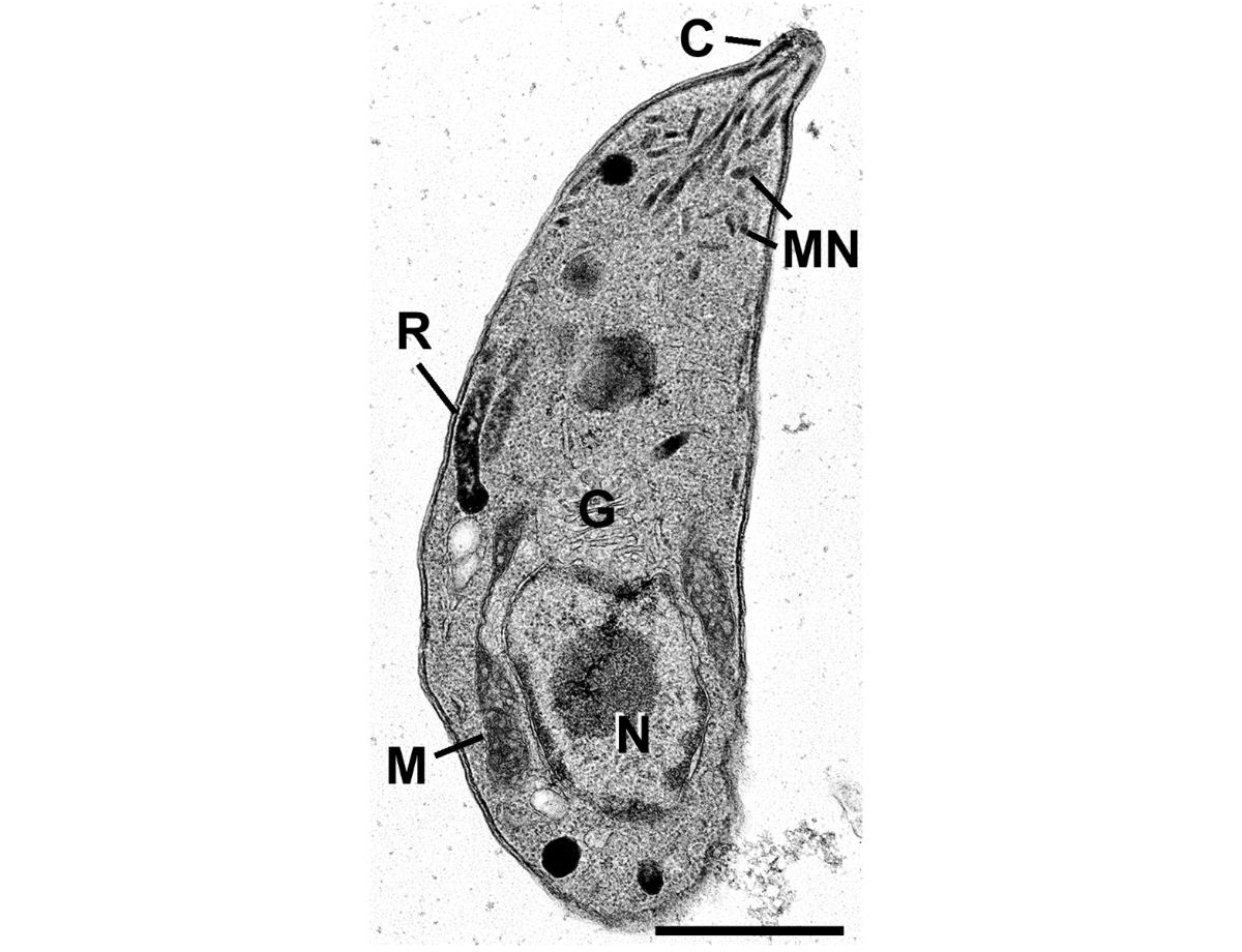
Figure 2
Extracellular tachyzoite with a fully extruded
conoid (C,
conoid; G, Golgi body; M, micronemes; Mi, mitochondrion; Nu, nucleus; R, rhoptry).
The apical complex is used by the parasite to penetrate the
host cell. Electron microscopy, scale bar = 1 μm. (Courtesy to Prof. David Ferguson,
Nuffield Department of Clinical Laboratory Science, University of Oxford, John Radcliffe
Hospital, Oxford, United Kingdom. Source: Koreny L, Zeeshan M, Barylyuk K, Tromer
EC, van Hooff JJE, Brady D, et al. Molecular characterization of the conoid complex
in Toxoplasma reveals its conservation in all apicomplexans, including Plasmodium
species. PLoS Biol. 2021 Mar 11;19(3):e3001081. doi: 10.1371/journal.pbio.3001081.)
Once in the bloodstream, the parasite spreads
throughout the host’s body. Upon reaching tissues, it becomes less active and
forms tissue cysts (bradyzoites), particularly in the skeletal muscles, heart, central
nervous system, and retina. These tissue cysts result in chronic, latent
infections. Host immune responses, both humoral and cellular, cannot eradicate
the parasite but keep it in check, allowing it to persist unnoticed unless
immunosuppression triggers reactivation. Only in definitive hosts does the
parasite complete its sexual cycle, releasing new infectious oocysts through
feline faeces and restarting the cycle.
Clinical manifestations – who falls ill?
Despite the millions of people globally being
exposed to T. gondii, clinically significant infections are rare. In
immunocompetent individuals, primary infection is often asymptomatic or mild, occasionally
presenting with mononucleosis-like symptoms such as fever, peripheral
lymphadenopathy, hepatosplenomegaly, and fatigue [1, 2]. Cervical or nuchal
lymph node enlargement can persist for extended periods, prompting further
medical investigations. Similar presentations can arise from EBV, CMV, HIV, bartonellosis,
tularaemia, lymph node tuberculosis, and lymphoma. Severe cases in immunocompetent
patients are more common in South America due to different, more virulent Toxoplasma
strains [1, 3].
Ocular toxoplasmosis and recurrences
Toxoplasmosis may primarily affect the eye and
is the leading cause of posterior uveitis. Around 1.5–3% of primary T.
gondii infections result in ocular toxoplasmosis (OT). Initially, OT was
thought to result from congenital infection, but most cases are now believed to be
acquired postnatally [11]. It typically
presents as unilateral central retinal necrosis (figure 3). If it involves the
macula, the area of sharpest vision, significant visual impairment or blindness
may result. Persistent vitreous opacities, strabismus, cataracts, and glaucoma
can also occur [11, 12].
Four out of five affected individuals experience
recurrences, often within two years of the primary infection. Recurrence can
follow trauma, stress, or hormonal fluctuations [12]. The specific
immunological factors responsible remain unclear. Unlike VZV reactivation, ageing
alone is not sufficient to trigger T. gondii reactivation. This differs
from reactivation in immunocompromised patients, which can lead to severe, necrotising
Toxoplasma retinitis.
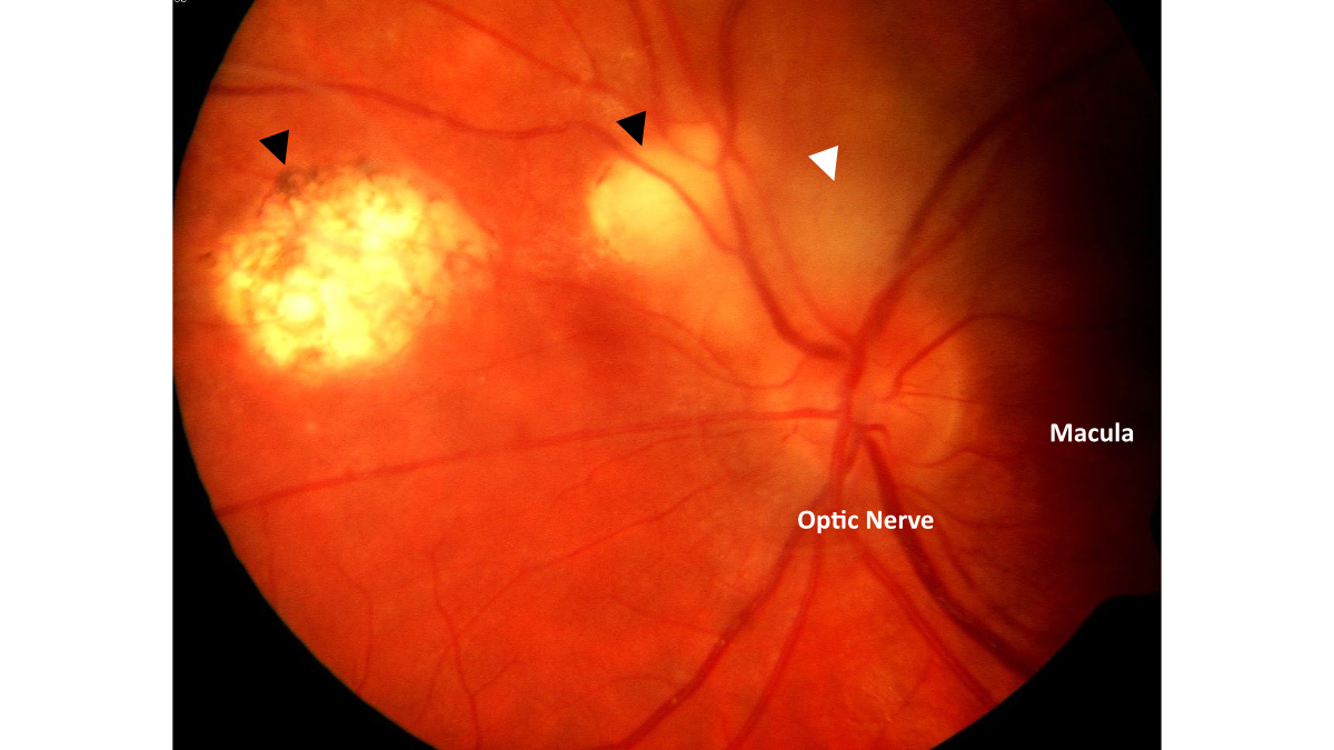
Figure 3
Recurrence of ocular toxoplasmosis
(discrete brightening and blurred disc margin, white arrow) nasally above the
disc. The black arrows point to old OT scars. (Courtesy to Prof. Dr. med. J. G.
Garweg, Bern Eye Clinic.)
Primary infection in pregnancy
Infection with T. gondii can also be acquired vertically through a primary
infection during pregnancy or shortly before conception. This feared infection
can, in the worst case, lead to miscarriage or permanent damage to the foetus.
However, the effects on the child are often less severe, and congenital
infection is fortunately rare. In 2022, 173 cases of congenital toxoplasmosis
were reported to the ECDC, corresponding to a rate of 5.8 per 100,000 live
births [13]. Of congenitally infected infants, 10–25% are symptomatic at birth.
The earlier the mother becomes infected, the lower the risk of foetal infection
(14%), but the more severe the potential damage to the child. The clinical
spectrum is broad, including early miscarriage, hydrocephalus, cerebral
calcifications, ocular lesions, hepatosplenomegaly, jaundice, and anaemia, among
others [14, 15]. By contrast, infection during the last trimester results in foetal
infection in nearly 60% of cases, though it tends to be milder. Some affected
children are not diagnosed until adolescence, when they present with impaired
vision, hearing or learning difficulties, or epilepsy.
Reactivation during immunosuppression
Reactivation of latent infection occurs when the
immune system is weakened and can no longer control the parasite. This can result
from highly immunosuppressive therapy, cellular immune dysfunction in haematological
cancers, or advanced untreated HIV infection (CD4 <100 cells/µl). In these
cases, the disease re-emerges as an opportunistic infection, causing severe
damage and potentially fatal outcomes without treatment. Reactivation can
manifest locally (e.g. in the eye, lungs, or CNS) or be disseminated. Patients
with encephalitis may exhibit altered consciousness, headaches, seizures, or
focal neurological deficits that correlate with multiple ring-enhancing lesions
seen on CT or MRI scans (figure 4). Severe, disseminated cases are observed
particularly in patients following allogeneic stem cell transplantation
(allo-HSCT) [1, 15]. In patients with HIV, these feared reactivations have
become rare with the advent of highly active antiretroviral therapy and
prophylactic treatment.
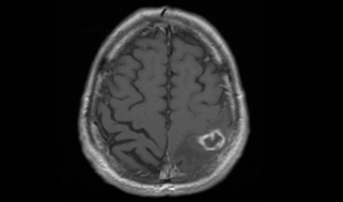
Figure 4
MRI of the neurocranium of a patient
with HIV and toxoplasmosis encephalitis. A centrally necrotic lesion is visible
in the left parietal region with ring enhancement. Axial T1 with contrast.
Toxoplasma gondii as a subtle manipulator?
In humans and rodents, T. gondii
primarily encysts in the CNS, prompting research into its effects on behaviour
and neurodegenerative diseases [16]. T. gondii appears to influence its
intermediate hosts, such as mice and small rodents, by reducing their ability
to detect predators, making them more prone to predation. This loss of predator
fear does not seem to be species-specific [16]. Other animals, such as wolves, also
show increased risk-taking behaviour following T. gondii infection.
While it remains unclear whether these effects benefit or harm the host, the
parasite undoubtedly benefits by facilitating its global spread.
The implications become unsettling when
considering research on T. gondii’s potential effects on humans. The
parasite may not simply lie dormant in host tissue without leaving traces.
Recent studies suggest that T. gondii might influence human personality
and decision-making [16–19]. Correlations have been reported between T.
gondii seroprevalence and authoritarian behaviour, tribalism, conservatism,
and risk-taking entrepreneurship [17, 18]. Furthermore, chronic CNS
inflammation caused by T. gondii is thought to contribute to psychiatric
or neurodegenerative diseases (e.g. schizophrenia, personality disorders, addiction,
and dementia) [18]. Possible underlying mechanisms include chronic
neuroinflammation; altered neurotransmitter metabolism, particularly dopamine; and
immune-mediated changes. Causality, however, remains unproven, and other
factors may contribute to a similar extent [16, 19]. While many questions
remain unanswered, it is plausible that T. gondii and other parasites
have shaped human evolution more profoundly than previously recognised.
Laboratory diagnosis
The Sabin-Feldman dye test, introduced in 1948, was
the first specific test to detect the parasite. It revealed the widespread
presence of T. gondii among warm-blooded animals [1, 15]. Today, various
immunoassays are available for serological diagnostics. Diagnosis is based on
the detection of specific IgM and IgG antibodies or on evidence of
seroconversion. IgM appears within 1–2 weeks and peaks at 4–8 weeks, whereas
IgG rises after 2–3 weeks, reaching maximal levels approximately 6–8 weeks after
infection [20]. Antibody levels decline following therapy or natural infection,
with IgG persisting lifelong, whereas IgM typically disappears within 6–9
months. However, IgM may persist for up to two years [20, 21], making it
challenging to distinguish recent infection from prior infection. IgG avidity
testing aids in this differentiation, as high avidity rules out infection
within the past 3–4 months [20].
Ocular toxoplasmosis diagnosis depends on the characteristic retinal
appearance and positive serology. Atypical presentations involving isolated eye
infection without detectable serum antibodies may require additional tests
(e.g. PCR of the aqueous or vitreous humour and antibody comparison between the
aqueous humour and serum) [11, 12]. Negative results from a single test should
not rule out the diagnosis, as test sensitivity varies.
Since infection during pregnancy can pose
a threat to the unborn child but is difficult to detect, several countries, particularly
France, have established surveillance programs with the goal of identifying maternal
seroconversion early and preventing transmission to the foetus [6, 13]. According
to the EU One Health 2023 Zoonoses report, six European countries, namely
Austria, Belgium, France, Greece, Slovakia, and Slovenia, had ongoing active
surveillance of congenital toxoplasmosis with mandatory screening of pregnant
women [13]. Over the past two decades, these resource-intensive programmes have
come under increasing criticism for the following reasons: cost constraints (e.g.
in settings with lower or declining prevalence), uncertainties about test
reliability, unwarranted anxiety and invasive diagnostics due to persistent IgM
antibodies, and insufficient evidence of clinical effectiveness [6, 15]. As a
result, many countries, including Switzerland, have discontinued or never
implemented routine screening programmes [6, 13].
Neonatal evaluation for toxoplasmosis at birth requires a great
deal of attention, as maternal infection is often asymptomatic. If congenital
infection is suspected, umbilical cord blood serology (IgA, IgM, IgG) is the
preferred method. PCR testing of cord blood or amniotic fluid is also possible,
though prenatal amniocentesis is discouraged due to the associated risks and
low PCR sensitivity (40–70%) [15].
Reactivation of latent infection in immunosuppressed T. gondii-positive patients
can be challenging. This applies particularly to patients following stem cell
transplantation (HSCT). In this setting, repeated qPCR testing of the blood has
become an established follow-up marker [15]. To confirm tissue infection in
suspected CNS toxoplasmosis or other focal infections, a biopsy for PCR and
histopathological analysis is often needed.
Treatment and pharmacologic prophylaxis
Toxoplasmosis in immunocompetent individuals is
typically self-limiting and does not require treatment. Therapy is necessary
for severe cases, disseminated infections, encephalitis, pneumonitis, myocarditis,
or chorioretinitis [15]. Since the 1950s, the gold standard for non-pregnant
patients has been the combination of pyrimethamine and sulfadiazine (Pyr-Sulf).
This combination inhibits folic acid metabolism, necessitating concurrent folic
acid supplementation. Alternatives include pyrimethamine combined with
clindamycin, azithromycin, dapsone, or the antimalarial drug atovaquone.
Co-trimoxazole, either alone or in combination with pyrimethamine or
clindamycin, also has supporting data. Corticosteroids may be indicated for
encephalitis with increased intracranial pressure or for chorioretinitis but
are otherwise not recommended [11, 15].
In early pregnancy (first trimester), the
macrolide spiramycin was traditionally the drug of choice because it does not
cross the placental barrier and is thought to prevent foetal transmission.
However, as mentioned above, many experts now discourage early treatment due to
a lack of efficacy [6, 15]. From the second trimester onwards and/or in cases
of suspected foetal infection, Pyr-Sulf alongside folic acid may be used [15]. It
should be noted that spiramycin is no longer available in the Swiss market, and
sulfadiazine must be imported. All therapeutic agents target the metabolically
active tachyzoite form of the parasite. Currently, no drugs can eliminate
dormant tissue cysts.
Severe infections in immunosuppressed patients
require prolonged treatment (>6 weeks), ideally extending for 1–2 weeks
beyond symptom resolution. In cases of persistent immunodeficiency, patients
need prophylaxis after therapy. Co-trimoxazole (TMP-SMX) remains the standard prophylaxis
for Toxoplasma gondii in severely immunosuppressed patients, including
those with advanced HIV, organ or stem-cell transplantation, or intensive
immunosuppressive therapy, while also providing concurrent protection against Pneumocystis
jirovecii [15].
Preventive measures and novel approaches
Avoiding toxoplasmosis entirely may seem almost
impossible given the parasite’s efficient spread and its robust oocysts. T.
gondii is nearly ubiquitous, found in food, water, and the environment [1, 10].
Nevertheless, seropositivity correlates with sociocultural factors, dietary
habits, and access to adequate sanitation and clean drinking water [1]. Safe
food, water, and hygiene practices can reduce the risk of infection. Key measures
include thoroughly cooking meat, washing fruits and vegetables, and maintaining
good hand hygiene after handling raw meat, soil, and cat litter.
Developing new therapeutic and preventive
strategies to protect the global population from this cunning parasite remains
a significant challenge. One Health research initiatives aim to create a
multisectoral vaccine to protect both humans and livestock while reducing
oocyst excretion by cats [22, 23]. Conditions are favourable for such advances.
For instance, an attenuated live vaccine already prevents miscarriages in
sheep. However, key challenges remain, particularly the parasite’s complex biology,
poor cross-species protection, limited translatability of animal models, and
persistent regulatory and economic obstacles [22, 23]. Another promising
approach involves therapeutics that prevent the parasite from invading host
cells and agents that are capable of eliminating tissue cysts [3, 24, 25, 26].
If these initiatives succeed, the world could be relieved of a significant
burden.
Sabine Majer, MD
Department of Infectious Diseases and Hospital Hygiene
Spital Thurgau AG
Kantonsspital Münsterlingen
Spitalcampus 1
CH-8596 Münsterlingen
sabine.majer[at]stgag.ch
References
1. Tenter AM, Heckeroth AR, Weiss LM. Toxoplasma gondii: From animals to humans. International
Journal for Parasitology, 2000;30(12–13,1217–1258
2. Farhab M, Waqar Aziz M, Shaukat A, Cao MX, Huang SY, Li L, Yuan YG. Review of Toxoplasmosis:
What we still need to do. Vet Sci 2025;12(8, 772
3. Human impact on the diversity and virulence of theubiquitous zoonotic parasite Toxoplasma
gondiiE. Shwab K, Saraf P, Zhub XQ, Zhou DH, McFerrin BM, Ajzenbergd D, Schares G,
Hammond-Aryee K, van Helden P, Higgins SA, Gerhold RW, Rosenthal BM, Zhaoc X, Dubey
JP, Su C. Human impact on the diversity and virulence of the ubiquitous zoonotic parasite
Toxoplasma gondii. PNAS 2018 Jul 2;115(29:E6956–E6963.
4. Sengupta PP, Jacob SS, Suresh KP, Rajamani S, Maharana SM. Exploring global trends
in human toxoplasmosis seroprevalence by meta-analysis. Exp Parasitol. 2025 Aug;275:108971.
doi: https://doi.org/10.1016/j.exppara.2025.108971
5. Calero-Bernal R, Gennari SM, Cano S, Salas-Fajardo MY, Ríos A, Álvarez-García G, Ortega-Mora LM.
Anti-Toxoplasma gondii antibodies in european residents: A systematic review and meta-analysis
of studies published between 2000 and 2020. Pathogens 2023;12(12:143
6. Swiss working group on congenital Toxoplasmosis. Rudin C, Boubaker K, Raeber PA, Vaudaux
B, Bucher HC, Garweg JG, Hoesli I, Kind C, Hohlfeld P. Toxoplasmosis during pregnancy
and infancy. A new approach for Switzerland. Swiss Medical Weekly, 2008 Dec 13,138(Suppl168:1–8
7. Lentini G, Ben Chaabene R, Vadas O, Ramakrishnan C, Mukherjee B, Mehta V, et al. Structural
insights into an atypical secretory pathway kinase crucial for Toxoplasma gondii invasion.
Nat Commun. 2021 Jun;12(1):3788. doi: https://doi.org/10.1038/s41467-021-24083-y
8. Torrey E, RH Yolken RH. Toxoplasma oocysts as a public health problem. Trends in Parasitology
August 2013,29(8:380-384 doi: https://doi.org/10.1016/j.pt.2013.06.001
9. Shapiro K, Bahia-Oliveira L, Dixon B, Dumètre A, de Wit LA, VanWormer E, et al. Environmental
transmission of Toxoplasma gondii: oocysts in water, soil and food. Food Waterborne Parasitol. 2019 Apr;15:e00049.
doi: https://doi.org/10.1016/j.fawpar.2019.e00049
10. Koutsoumanis K, Allende A, Alvarez-Ordóñez A, Bolton D, Bover-Cid S, Chemaly M, et
al.; EFSA Panel on Biological Hazards (BIOHAZ). Public health risks associated with
food-borne parasites. EFSA J. 2018 Dec;16(12):e05495. 10.2903/j.efsa.2018.5495
11. Kalogeropoulos D, Sakkas H, Mohammed B, Vartholomatos G, Malamos K, Sreekantam S,
Kanavaros P, Kalogeropoulos C. Ocular toxoplasmosis: A review of the current diagnostic
and therapeutic approaches. Int Ophthalmol. 2022;42(1:295–321 doi: https://doi.org/10.1007/s10792-021-01994-9
12. Cifuentes-González C, Rojas-Carabali W, Olate Pérez A, Carvalho E, Valenzuela F, Miguel-Escuder L,
Ormaechea M, Heredia M, Baquero-Ospina P, Adan A, Curi A, Schlaen A, Urzua CA, Couto C,
Arellanes L, de-la-Torre A. Risk factors for recurrences and visual impairment in
patients with ocular toxoplasmosis: A systematic review and meta-analysis. PLoS One.
2023 Apr 3;18(4:e0283845
13. European Food Safety Authority (EFSA); European Centre for Disease Prevention and
Control (ECDC). The European Union One Health 2023 Zoonoses report. EFSA J. 2024 Dec;22(12):e9106.
14. Dubey JP, Murata FHA, Cerqueira-Cézar CK, Kwok OCH, Villena I. Congenital toxoplasmosis
in humans: An update of worldwide rate of congenital infections. Parasitology. 2021 Jun 18;148(12:1406–1416
15. Dunay IR, Gajurel K, Dhakal R, Liesenfeld O, Montoya JG. Treatment of Toxoplasmosis:
Historical perspective, animal models, and current clinical practice. Clin Microbiol
Rev. 2018 Sep 12;31(4:e00057-17
16. Johnson HJ, Koshy AA. Latent Toxoplasmosis effects on rodents and humans: How much
is real and how much is media hype? mBio 2020 Mar,17;11(2
17. J Flegr. How and why Toxoplasma makes us crazy. Trends in Parasitology. 2013 April,29(4:156-163
doi: https://doi.org/10.1016/j.pt.2013.01.007
18. Kopecky R, Příplatová L, Boschetti S, Talmont-Kaminski K, Flegr J. Le petit Machiavellian
prince: Effects of latent Toxoplasmosis on political beliefs and values. Evol Psychol.
2022 Jul 29;20(3:14747049221112657
19. de Haan L, Sutterland AL, Schotborgh JV, Schirmbeck F, de Haan L. Association of Toxoplasma
gondii seropositivity with cognitive function in healthy people. A systematic review
and meta-analysis. JAMA Psychiatry. 2021 Oct 1;78(10:1103-1112
20. Teimouri A, Mohtasebi S, Kazemirad E, Keshavarz H. Role of Toxoplasma gondii IgG Avidity
testing in discriminating between acute and chronic Toxoplasmosis in pregnancy. J
Clin Microbiol. 2020 Aug 24;58(9:e00505-20
21. Gras L, Gilbert RE, Wallon M, Peyron F, Cortina-Borja M. Duration of the IgM response
in women acquiring Toxoplasma gondii during pregnancy: implications for clinical practice
and cross-sectional incidence studies. Epidemiology & Infection. June 2004; 132(3:541-548
doi: https://doi.org/10.1017/S0950268803001948
22. Innes EA, Hamilton C, Garcia JL, Chryssafidis A, Smith D. A one health approach to
vaccines against Toxoplasma gondii. Food Waterborne Parasitol. 2019 Apr;15:e00053. doi: https://doi.org/10.1016/j.fawpar.2019.e00053
23. Zhang Y, Li D, Lu S, Zheng B. Toxoplasmosis vaccines: what we have and where to go? NPJ
Vaccines. 2022 Oct;7(1):131. doi: https://doi.org/10.1038/s41541-022-00563-0
24. Smith NC, Goulart C, Hayward JA, Kupz A, Miller CM, van Dooren GG. Control of human
toxoplasmosis. Review Article. International Journal for Parasitology. Febr. 2021;51:95–121.
25. da Silva M, Teixeira C, Gomes P, Borges M. Promising drug targets and compounds with
anti-Toxoplasma gondii activity. Microorganisms. 2021 Sep 15;9(9:1960
26 El Hajj R, Tawk L, Itani S, Hamie M, Ezzeddine J, El Sabban M, El Hajj H. Toxoplasmosis:
Current and emerging parasite druggable targets. Microorganisms. 2021 Dec 7;9(12:2531