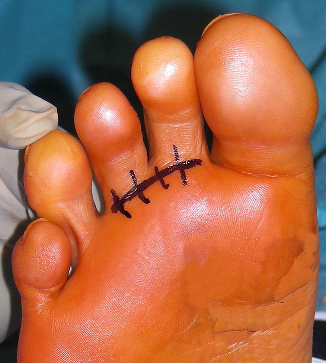
Figure 1 Distal transverse plantar incision to the second web space.
DOI: https://doi.org/10.4414/smw.2020.20347
Operative treatment of Morton’s neuroma is indicated after failure of conservative management [1]. Neurectomy is the most common procedure, involving resection of the affected nerve [2, 3]. Neurolysis is an alternative treatment and consists of the release of the transverse intermetatarsal ligament without nerve resection. This procedure can be open, percutaneous or endoscopic [4–6].
Neurectomy can be performed through longitudinal dorsal [7], web-splitting [8], longitudinal plantar [9], and distal transverse plantar [10] approaches. Because of its anatomical location, a plantar approach provides optimal exposure of the nerve. However, painful scar formation is regarded as a potential drawback associated with the longitudinal plantar approach [11, 12]. This can be avoided by using a longitudinal dorsal approach. Nevertheless, the dorsal approach has some disadvantages, including the need for transection of the transverse metatarsal ligament and a more difficult visualisation of the nerve, and is associated with a higher risk of stump neuroma or missed neuroma [13, 14]. On the other hand, the distal transverse plantar approach may provide optimal exposure of the nerve with a lower complication rate related to the scar formation.
The main purpose of this study was to evaluate the long-term clinical results following a distal transverse plantar approach in the operative treatment of Morton’s neuroma. The secondary purpose was to compare the results between patients who had undergone neurectomy alone and patients in whom additional procedures for the treatment of concomitant foot disorders were performed.
After obtaining approval of the local ethics committee for the study, we identified all cases of patients who underwent primary excision of a Morton’s neuroma through a distal transverse plantar approach at our institution between 2004 and 2011 by a single surgeon (senior author, XC) or under his direct supervision. Only patients with available histopathological confirmation of the diagnosis were selected. Each patient was then invited to take part in the study. The patients who agreed to participate were included after giving their informed consent. Of a total of 69 operated patients, we identified 65 in whom histopathological confirmation of the diagnosis was available. From those, 49 patients (45 women and 4 men) agreed to be included in the study and signed the informed consent form. Reasons for non-inclusion in 16 patients were: no response to a telephone call and mailing in 9 patients, refusal to attend for clinical examination because of remote residence in 5 patients, and death in 2 cases. Operative reports were reviewed for the exact procedure that had been performed. Charts were screened for the postoperative course, including the initial healing of the scar and the need for revision surgery. Patients were assessed clinically after a median of 7.9 years (range 4–12) postoperatively by one of the two independent observers (THNN and HH) using two validated outcome scores, the Foot and Ankle Ability Measure (FAAM) [15] and the Vancouver Scar Scale (VSS) [16].
The Foot and Ankle Ability Measure (FAAM) is a 29-item questionnaire developed to assess the physical function of individuals with foot and ankle disorders. This self-report outcome instrument is available in English, German, French and Persian. Based on the requirements of our study demographics, we opted for the validated French version of the FAAM [17]. The questionnaire is divided in two sub-scores: the activities of daily living (ADL) sub-score (21-item), and the sports sub-score (8-item). Each item is scored on a 5-point Likert scale (4 to 0) from “no difficulty at all” to “unable to do”. Item score totals, which range from 0 to 84 for the ADL sub-score and 0 to 32 for the sports sub-score, are transformed into percentage scores. Higher scores represent higher levels of function for each sub-score, with 100% representing normal functional status.
The Vancouver Scar Scale (VSS) is a validated clinical assessment of scars introduced in 1990 by Sullivan [16]. It is probably the most widely known scar scale [18]. Initially developed for burn scars, it has been later applied to linear scars and has shown acceptable internal consistency, despite a poor-to-moderate interobserver reliability [19, 20]. We used a modified version of the VSS as described by Baryza et al [21]. This version includes a 15-point scale instead of the original 13-point scale and we decided to use it for a more subtle evaluation. It is based on four parameters: pigmentation, vascularity, height and pliability. Each parameter has its own set of scores: 0 to 3 for pigmentation (normal, hypopigmented, mixed, hyperpigmented), 0 to 3 for the vascularity (normal, pink, red, purple), 0 to 4 for height (flat, > 0 to 1 mm, > 1 to 2 mm, > 2 to 4 mm, > 4 mm) and 0 to 5 for pliability (normal, supple, yielding, firm, ropes, contracture). The total score ranges from 0 to 15, a score of 0 representing normal skin.
Additionally, while scoring the scar according to the VSS, one of the two independent observers (THNN and HH) examined the scar for painful palpation.
Moreover, the patients were asked if they would accept the operative treatment again if needed, and if they would recommend it to a friend.
All operations were performed by a senior surgeon (XC) or under his direct supervision. A tourniquet, inflated at 300 mm Hg, was applied to the thigh in the case of general anaesthesia or to the supramalleolar portion of the leg in the case of peripheral anaesthesia (popliteal block). Patients were positioned supine and a transverse plantar incision was made distal to the metatarsal heads (fig. 1). In all cases, firm plantar pressure was applied under the metatarsal heads in order to exclude any overlap between the incision and the weight bearing surface of the forefoot (fig. 2). After exposure of the subcutaneous tissues, the neuroma was identified (fig. 3) and a Y resection of the digital nerve was carried out (fig. 4). The tourniquet was released, haemostasis was achieved, and the skin was closed with absorbable sutures. Weight bearing as tolerated was allowed from day one following surgery, with use of a special post-operative shoe (DARCO MedSurg™) for 3–4 weeks. The dressing was changed once a week until healing occurred.
Figure 1 Distal transverse plantar incision to the second web space.
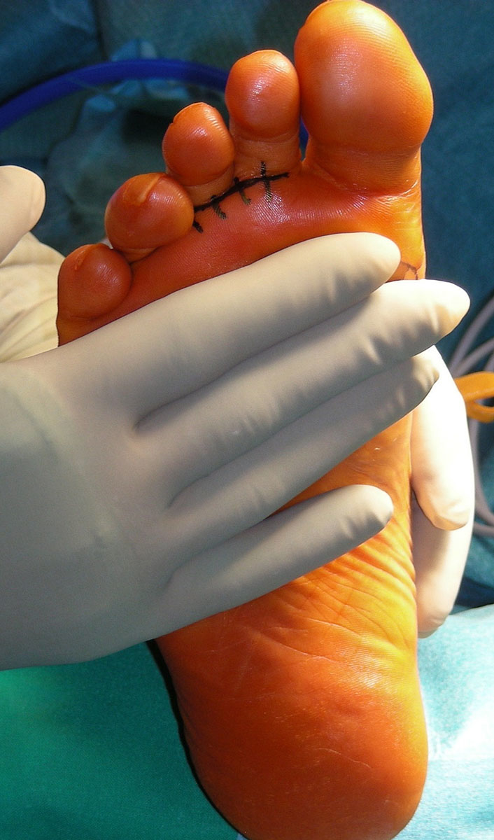
Figure 2 Plantar pressure is applied to the forefoot in order to exclude any overlap between the incision and the weight bearing surface of the forefoot.

Figure 3 After exposure of the subcutaneous tissues, the neuroma is identified, here in the second web space.
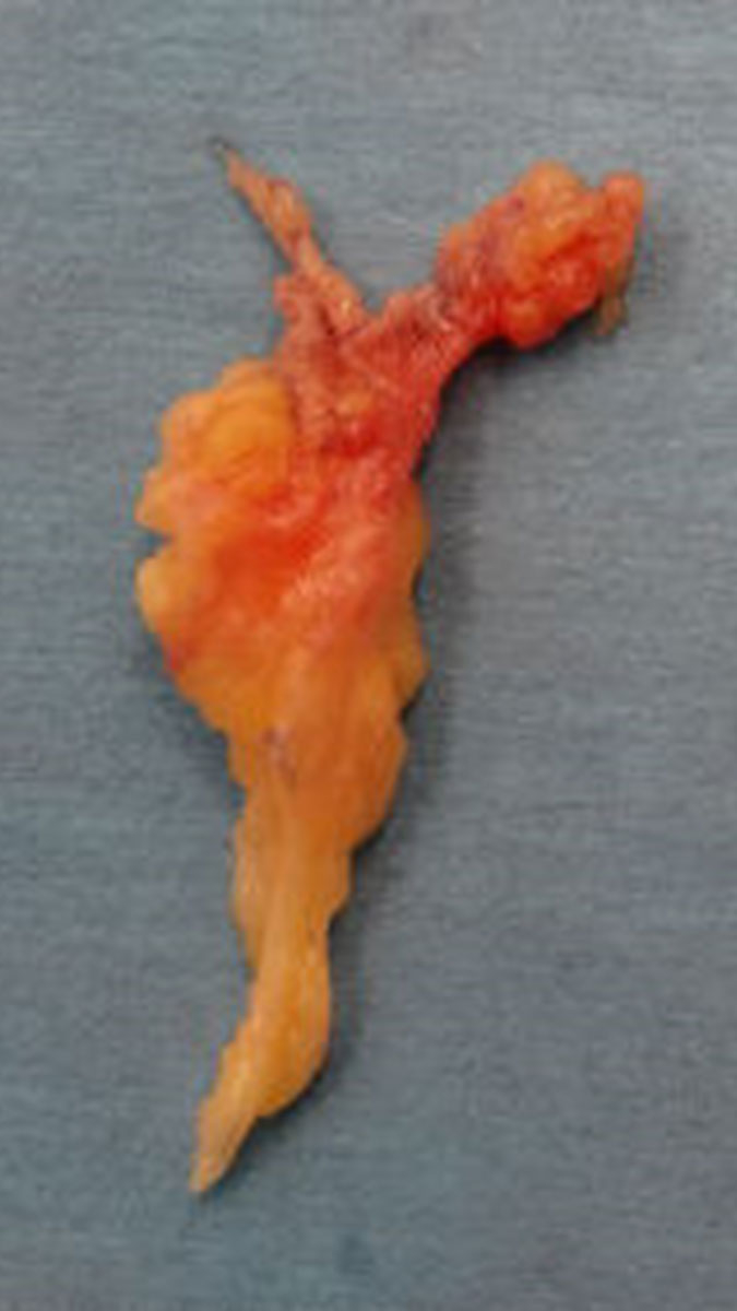
Figure 4 A Y resection of the digital nerve is carried out and the sample is sent for histopathological confirmation of the diagnosis.
Outcomes from the two questionnaires are reported as mean and standard deviation. The unit of analysis was per foot. The outcomes of patients operated on for neurectomy alone and those having a concomitant procedure were compared using an independent Student’s-t-test. A chi-square test was used to compare the rates of recommendation, and acceptance of redoing the procedure – between patients with and without additional operative procedures. The significance was set at p <0.05.
The median age at the time of surgery was 59 years (range 24–77). In total, 103 Morton’s neuromas were excised from 58 feet belonging to the 49 patients. Both feet were involved in 9 patients, the right foot in 27 patients and the left foot in 31 patients. A single intermetatarsal space was involved in 11 feet (3 with the second intermetatarsal space, 8 with the third intermetatarsal space). Both the second and third intermetatarsal spaces were involved in 45 feet, and the third and fourth intermetatarsal spaces were involved in 2 feet (table 1). Concurrent foot surgery was performed in 30 patients. These procedures are listed in table 2.
Table 1 Demographics.
| Sex (n): | |
| – Male | 4 |
| – Female | 45 |
| Age (years), mean (range) | 59 (24–77) |
| Side (n): | |
| – Right | 27 |
| – Left | 31 |
| – Bilateral | 9 |
| Webspace(s) (n): | |
| – 2nd | 3 |
| – 3rd | 8 |
| – 2nd and 3rd | 45 |
| – 3rd and 4th | 2 |
Table 2 List of cases with additional procedures.
| Additional procedures | No. of patients |
|---|---|
| Hallux valgus correction | 14 |
| Lesser metatarsal osteotomies | 3 |
| Combined hallux valgus correction and lesser metatarsal osteotomies | 11 |
| Other procedures | 6 |
The mean FAAM score was 84.8% (range 16–100%). The mean ADL and sports sub-scores were 89.6% (range 30–100%) and 80.0% (range 4–100%), respectively. The mean VSS was 1.57 (range 0–6). Figure 5 shows representative scars at a mean of 7.9 years follow up. If needed, 86% of patients would have the operative treatment again (REDO) and 84% would recommend it to a friend (RECOMM). In 8 patients, clinical examination revealed a tender scar on palpation although no scar-related pain while walking was reported. Among these eight patients, all except one would have had the surgery redone and would recommend it to a friend.
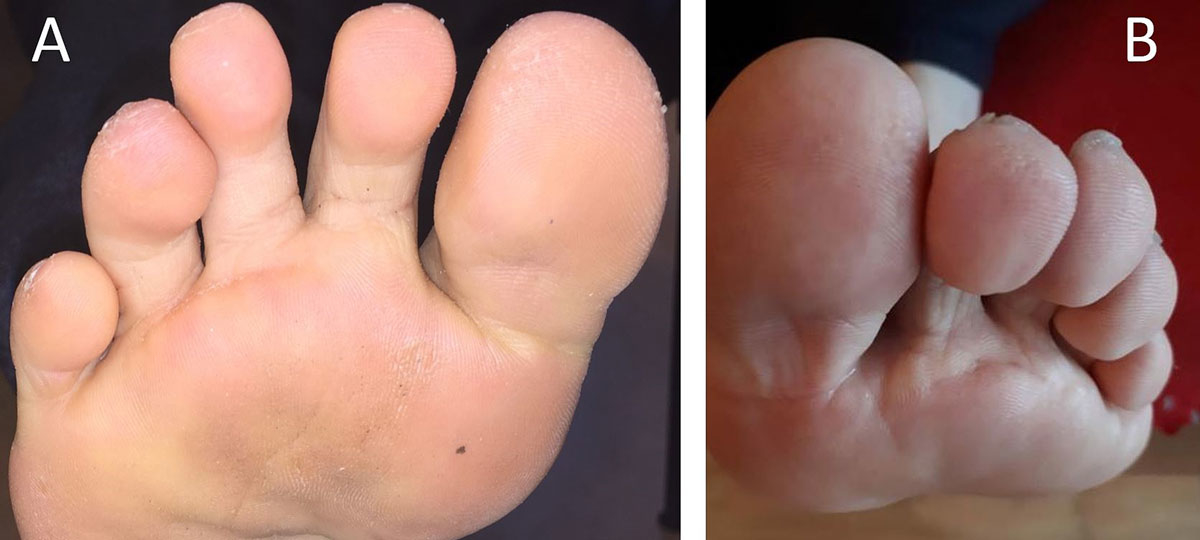
Figure 5 Representative scar samples at a mean of 7.9 years follow up in two different patients. A third web space, B second web space.
Comparison between patients who had undergone neurectomy alone (group 1) and patients in whom additional procedures for the treatment of concomitant foot disorders were performed (group 2) revealed a significantly better outcome in group 1 for FAAM total score (p = 0.0007), FAAM ADL (p = 0.02) and sports (p = 0.0003) sub-scores as well as for the RECOMM for the procedure (p = 0.04). No significant difference between the groups were reported for the VSS score and REDO (table 3).
Table 3 Comparison between patients who had undergone neurectomy alone (group 1) and patients in whom additional procedures for the treatment of concomitant foot disorders was performed (group 2).
| Outcome measures |
Group 1
(n = 19) |
Group 2
(n = 30) |
p-value |
|---|---|---|---|
| FAAM total | 88.12 ± 23% | 65.94 ± 23% | 0.00007 |
| FAAM ADL | 95.76 ± 12% | 85.24 ± 18% | 0.02 |
| FAAM sports | 96.56 ± 8% | 66.08 ± 28% | 0.0003 |
| VSS | 1.21 ± 1.61 | 1.82 ± 1.78 | NS |
| REDO | 96% | 79% | NS |
| RECOMM | 96% | 76% | 0.04 |
| ADL = activities of daily life; FAAM = Foot and Ankle Ability Measure (FAAM); RECOMM = patient would recommend the procedure to a friend; REDO = patient would undergo the procedure again if necessary; VSS = Vancouver Scar Scale | |||
We observed no complication that required an additional procedure. However, in five patients delayed scar healing of more than three weeks was noted.
The present study demonstrates the efficacy of the distal transverse plantar approach in the treatment of Morton’s neuroma, confirmed by the objective results obtained from two validated outcome scales, the FAAM (Foot and Ankle Ability Measure) and the VSS (Vancouver Scar Scale). Patient’s scores reached 89.6% for the FAAM ADL sub-score, 80.0% for the FAAM sports sub-score and 1.57 points for the VSS score. Painful palpation of the scar was detected during clinical examination in eight patients, of whom seven reported no negative effect on function with the procedure. At follow-up we observed no recurrence and a very low complication rate. Notably, performing neurectomy alone resulted in a better outcome than when in combination with additional foot surgery. Simultaneous involvement of the 2nd and 3rd web spaces in our study was higher than previously reported. This is explained by our diagnostic procedure, which relied on patient’s history and clinical examination only. The threshold for exploration of the adjacent web space was set very low: it was decided to do so if the patient’s symptoms and the sagittal palpation were consistent with the involvement of two web spaces. Of course this practice may raise the concern that these patients suffered rather from a forefoot dysbalance or overload, and that unnecessary Morton’s neuroma surgery was performed. Histopathological examination, however, confirmed our clinical diagnosis and surgical decision in all cases even if, in 90% of cases, the degenerative changes were more marked in one of the two resected samples. The high scores obtained in our study, which included many patients in whom two adjacent web spaces were explored, illustrates one of the benefits of the transverse plantar approach, which allows access to two adjacent web spaces through a single incision.
The transverse plantar approach was described in 1950 by Kaplan [10], who aimed to avoid two disadvantages associated with the dorsal approach: the division of the deep transverse intermetatarsal ligament and the considerable depth of the exposure. Kaplan placed the incision at the root of the toes, slightly proximal to the interdigital folds. His observation of three cases showed that the exposure of the nerve was easier than through the dorsal approach, and that the incision did not interfere with weight bearing nor produce sloughing of the skin. Kaplan noted, however, that the healing of the wound was slightly longer than after a dorsal approach. Studies evaluating the transverse plantar approach are sparse. A retrospective long-term follow-up study of 160 patients was conducted by Nery et al. [22]. In less than one third (30%) of their patients they performed additional procedures for concomitant disorders, compared with less than two thirds (61%) of patients in the present study. They reported good results in 143 patients (89.4%), fair results in 11 patients (6.9%) and poor results in 6 patients (3.8%). Patients with poor results suffered from pain and paraesthesia, were re-operated on for recurrent neuroma and then became asymptomatic. The main reason for a fair result was the presence of scar symptoms (5% of the study group), including skin hardening and discomfort when wearing high heeled shoes. The authors concluded that the results of the Morton’s excision through a transverse plantar approach are at least comparable to those of studies evaluating different operative approaches and that the recurrence rate is low (3.8%). In a retrospective comparative study of 44 patients, 15 who underwent a transverse plantar and 29 who underwent a longitudinal dorsal approach, Wilson and Kuwada reported a complete relief of symptoms in 100% (transverse plantar) and 68% (longitudinal dorsal) of patients [23]. In the transverse plantar group, two patients suffered from a painful scar (13%) and two other from wound dehiscence. No stump neuroma was observed. In the longitudinal dorsal group, six stump neuromas (21%), two painful scars (6.9%), one deep vein thrombosis and one haematoma were reported. The authors concluded that the transverse plantar approach is a viable primary choice for neuroma excision because it provides excellent anatomic exposure and easy access to the neuroma, with no stump neuroma reported. Like Wilson and Kuwada [23], we observed no recurrence. Furthermore, delayed scar healing in five patients is consistent with the observations of Kaplan [10] and similar to Wilson and Kuwada’s results [23].
An original aspect of our study, compared with the aforementioned studies, is that we were able to objectify the good results of the transverse plantar approach using validated outcome scales. In particular, the VSS provides a clear statement about the long-term scar outcome. The low VSS scores observed in the present study demonstrate the safety of the transverse plantar approach in terms of scar healing.
The present study was not designed to compare different approaches. However, our results seem not to be inferior to those obtained by longitudinal dorsal or plantar approaches [11–14, 24]. Of note is the absence of recurrent or stump neuromas in our series, which may be explained by the excellent visualisation of the nerve as already emphasised in previous reports [10, 22, 23].
The strength of our study is the use, for the first time to our knowledge, of two validated outcome scores – the FAAM and the VSS – to confirm the good long-term results obtained in previous studies on the transverse plantar approach in the treatment of Morton’s neuroma. There are, however, three limitations in the present study: its retrospective design, and, therefore, the absence of baseline data of the scales used for assessment; the fact that 61% of patients had concomitant surgery of the forefoot; and the non-inclusion of 16 patients for missing data. The retrospective design is a limitation that we cannot ignore, but we consider it to be acceptable as the study focused solely on the long-term outcome of a single technique. Nevertheless, we regret especially that monitoring of scar healing was not possible. Future prospective studies comparing different surgical approaches are recommended to quantify early postoperative recovery, which could further help in clinical decision making with respect to time to return to work and/or sports. The rate of concomitant surgery in our series is higher than that reported by Nery et al. [22]. However, to perform simultaneously additional procedures if required is frequently a clinical necessity. Furthermore, including these patients adds value to the study because it allowed a comparison of outcome between neurectomy performed alone and in combination with further forefoot surgery. With regard to the missing data, it is unfortunately inherent in most retrospective studies.
There is currently no consensus on the ideal approach for the operative treatment of Morton’s neuromas. Our results are comparable to those of studies that have evaluated the same operative approach or a different one. The transverse plantar approach results in good objective outcome scores, including scar healing, at long-term. This is our preferred technique because, in our experience, it offers optimal visualisation of the nerve, does not require deep dissection, and allows the exposure of two adjacent web spaces of the foot through a single incision.
No financial support and no other potential conflict of interest relevant to this article was reported.
1 Thomas JL , Blitch EL, 4th , Chaney DM , Dinucci KA , Eickmeier K , Rubin LG , et al.; Clinical Practice Guideline Forefoot Disorders Panel. Diagnosis and treatment of forefoot disorders. Section 3. Morton’s intermetatarsal neuroma. J Foot Ankle Surg. 2009;48(2):251–6. doi:.https://doi.org/10.1053/j.jfas.2008.12.005
2 Bennett GL , Graham CE , Mauldin DM . Morton’s interdigital neuroma: a comprehensive treatment protocol. Foot Ankle Int. 1995;16(12):760–3. doi:.https://doi.org/10.1177/107110079501601204
3 Mann RA , Reynolds JC . Interdigital neuroma--a critical clinical analysis. Foot Ankle. 1983;3(4):238–43. doi:.https://doi.org/10.1177/107110078300300411
4 Barrett SL , Pignetti TT . Endoscopic decompression for intermetatarsal nerve entrapment--the EDIN technique: preliminary study with cadaveric specimens; early clinical results. J Foot Ankle Surg. 1994;33(5):503–8.
5 Finney W , Wiener SN , Catanzariti F . Treatment of Morton’s neuroma using percutaneous electrocoagulation. J Am Podiatr Med Assoc. 1989;79(12):615–8. doi:.https://doi.org/10.7547/87507315-79-12-615
6 Gauthier G . Thomas Morton’s disease: a nerve entrapment syndrome. A new surgical technique. Clin Orthop Relat Res. 1979;(142):90–2.
7 McKEEVER DC . Surgical approach for neuroma of plantar digital nerve (Morton’s metatarsalgia). J Bone Joint Surg Am. 1952;34(2):490. doi:.https://doi.org/10.2106/00004623-195234020-00019
8 McElvenny R . The etiology and surgical treatment of intractable pain about the fourth metatarsophalangeal joint (Morton’s Toe). J Bone Joint Surg. 1943;25:675–9.
9 Betts LO . Morton’s metatarsalgia: Neuritis of the fourth digital nerve. Med J Aust. 1940;1(15):514–5. doi:.https://doi.org/10.5694/j.1326-5377.1940.tb46191.x
10 Kaplan EB . Surgical approach to the plantar digital nerves. Bull Hosp Jt Dis. 1950;11(1):96–7.
11 Akermark C , Saartok T , Zuber Z . A prospective 2-year follow-up study of plantar incisions in the treatment of primary intermetatarsal neuromas (Morton’s neuroma). Foot Ankle Surg. 2008;14(2):67–73. doi:.https://doi.org/10.1016/j.fas.2007.10.004
12 Kundert HP , Plaass C , Stukenborg-Colsman C , Waizy H . Excision of Morton’s Neuroma Using a Longitudinal Plantar Approach: A Midterm Follow-up Study. Foot Ankle Spec. 2016;9(1):37–42. doi:.https://doi.org/10.1177/1938640015599032
13 Akermark C , Crone H , Skoog A , Weidenhielm L . A prospective randomized controlled trial of plantar versus dorsal incisions for operative treatment of primary Morton’s neuroma. Foot Ankle Int. 2013;34(9):1198–204. doi:.https://doi.org/10.1177/1071100713484300
14 Bucknall V , Rutherford D , MacDonald D , Shalaby H , McKinley J , Breusch SJ . Outcomes following excision of Morton’s interdigital neuroma: a prospective study. Bone Joint J. 2016;98-B(10):1376–81. doi:.https://doi.org/10.1302/0301-620X.98B10.37610
15 Martin RL , Irrgang JJ , Burdett RG , Conti SF , Van Swearingen JM . Evidence of validity for the Foot and Ankle Ability Measure (FAAM). Foot Ankle Int. 2005;26(11):968–83. doi:.https://doi.org/10.1177/107110070502601113
16 Sullivan T , Smith J , Kermode J , McIver E , Courtemanche DJ . Rating the burn scar. J Burn Care Rehabil. 1990;11(3):256–60. doi:.https://doi.org/10.1097/00004630-199005000-00014
17 Borloz S , Crevoisier X , Deriaz O , Ballabeni P , Martin RL , Luthi F . Evidence for validity and reliability of a French version of the FAAM. BMC Musculoskelet Disord. 2011;12(1):40. doi:.https://doi.org/10.1186/1471-2474-12-40
18 Vercelli S , Ferriero G , Sartorio F , Cisari C , Bravini E . Clinimetric properties and clinical utility in rehabilitation of postsurgical scar rating scales: a systematic review. Int J Rehabil Res. 2015;38(4):279–86. doi:.https://doi.org/10.1097/MRR.0000000000000134
19 Truong PT , Abnousi F , Yong CM , Hayashi A , Runkel JA , Phillips T , et al. Standardized assessment of breast cancer surgical scars integrating the Vancouver Scar Scale, Short-Form McGill Pain Questionnaire, and patients’ perspectives. Plast Reconstr Surg. 2005;116(5):1291–9. doi:.https://doi.org/10.1097/01.prs.0000181520.87883.94
20 Nicholas RS , Falvey H , Lemonas P , Damodaran G , Ghanem AM , Selim F , et al. Patient-related keloid scar assessment and outcome measures. Plast Reconstr Surg. 2012;129(3):648–56. doi:.https://doi.org/10.1097/PRS.0b013e3182402c51
21 Baryza MJ , Baryza GA . The Vancouver Scar Scale: an administration tool and its interrater reliability. J Burn Care Rehabil. 1995;16(5):535–8. doi:.https://doi.org/10.1097/00004630-199509000-00013
22 Nery C , Raduan F , Del Buono A , Asaumi ID , Maffulli N . Plantar approach for excision of a Morton neuroma: a long-term follow-up study. J Bone Joint Surg Am. 2012;94(7):654–8. doi:.https://doi.org/10.2106/JBJS.K.00122
23 Wilson S , Kuwada GT . Retrospective study of the use of a plantar transverse incision versus a dorsal incision for excision of neuroma. J Foot Ankle Surg. 1995;34(6):537–40. doi:.https://doi.org/10.1016/S1067-2516(09)80074-4
24 Coughlin MJ , Pinsonneault T . Operative treatment of interdigital neuroma. A long-term follow-up study. J Bone Joint Surg Am. 2001;83(9):1321–8. doi:.https://doi.org/10.2106/00004623-200109000-00005
No financial support and no other potential conflict of interest relevant to this article was reported.