
DOI: https://doi.org/10.4414/smw.2012.13597
Diesel is the fuel of choice for use in vehicles of mass transportation including lorries, buses, and trains. Combusted diesel exhaust (DE) is a complex mixture of gaseous and particulate compounds resulting from an incomplete combustion of diesel fuel. While over 100 substances have been identified in DE [1], major components include nitrogen and sulfur oxides, carbon monoxide (CO), organic compounds (including aromatic and alkane substances), and particles [2]. Relative to those using gasoline, diesel engines emit higher concentrations of nitrogen and sulfur oxides, particle numbers, and organic molecules [2]. Diesel exhaust particle (DEP) is usually <25% of the emission mass and composed of elemental carbon (50–75% of the mass depending on engine age, technology, fuel characteristics, and operating conditions), organics (19–43%), sulfates, nitrates, and metals. Approximately >95% of DEP is fine (PM2.5) while ultrafine particle can account for up to 20% of the total mass. The ultrafine component of DEP always constitutes the majority of the total particle number [3]; particles of this size present an enormous surface area allowing absorbance of compounds and subsequent transport into the distal portions of the lung.
DEP concentrations in ambient air are employed as a measure of exposure to DE. DEP mass in ambient PM is variable approximating zero in rural areas to 35% of PM10 in urban areas. The mean DEP exposure in the United States is calculated to be 2 μg/m3 but can be greater in both vehicles and the immediate vicinity of major streets and highways where it can approach 20–25 µg/m3[4–6]. Areas of significantly increased DE exposure along transportation avenues have been described with DEP concentrations >100 μg/m3 (up to a maximum exposure of approximately 350 ug/m3 with the duration being seconds) during simulations of a vehicle with a 2006 diesel engine driving by [7].
Recent changes in diesel engines have altered exhaust characteristics [8, 9]. Newer engines emit reduced CO, organics, and nitrogen oxides, [10]. In addition, ambient air DEP concentrations are lower now than in the past due to improvements in diesel-engine technology. While there is evidence that DEP mass is lower in the exhaust of newer engines, studies suggest that nanoparticle emissions may not be decreased.
Controlled human exposures refer to human investigation which places either healthy subjects or patients in a chamber into which an exogenous agent is introduced. Such studies have contributed to understanding pollutant-induced health effects and, relative to epidemiological and other forms of investigation, provide several advantages (table 1) [11, 12]. Subsequently, controlled human exposures have generated much knowledge about the underlying mechanisms of how air pollutants affect the body. There are also limitations of this methodology (table 1). While it is recognised that controlled human exposures employ particle concentrations which can be more relevant to ambient levels than those in animal studies, they frequently use doses which are high relative to ambient concentrations in order to increase detection of an effect while guaranteeing safety for the subject. In past investigations, DEP concentrations used in controlled human exposures approached one to two orders of magnitude greater than average environmental exposures of the general population (e.g., 300 vs. 3 µg/m3 respectively). Controlled human exposures often focus on a single air pollutant; a scenario that is rarely achieved in real life. The observed biological effects of a controlled human exposure are frequently small and the signal to noise ratio can be unfavourable. Short one-time exposures (which usually are a few hours) make it difficult to relate a “no effect” result to true environmental exposures which, in contrast, are both prolonged and repetitive. Study endpoints for controlled human exposures can frequently be limited to samples that are easily accessible, e.g., symptoms, examination, blood, urine, pulmonary function tests, electrocardiographic indices, sputum, breath condensate, and sometimes bronchoscopy specimens (bronchoalveolar lavage fluid, bronchial brushing and endobronchial biopsy). Finally, it is rather complex, if not impossible, to extrapolate the changes in endpoints obtained during controlled human exposures to those employed in epidemiological studies (e.g. mortality and hospital admissions for chronic obstructive pulmonary disease).
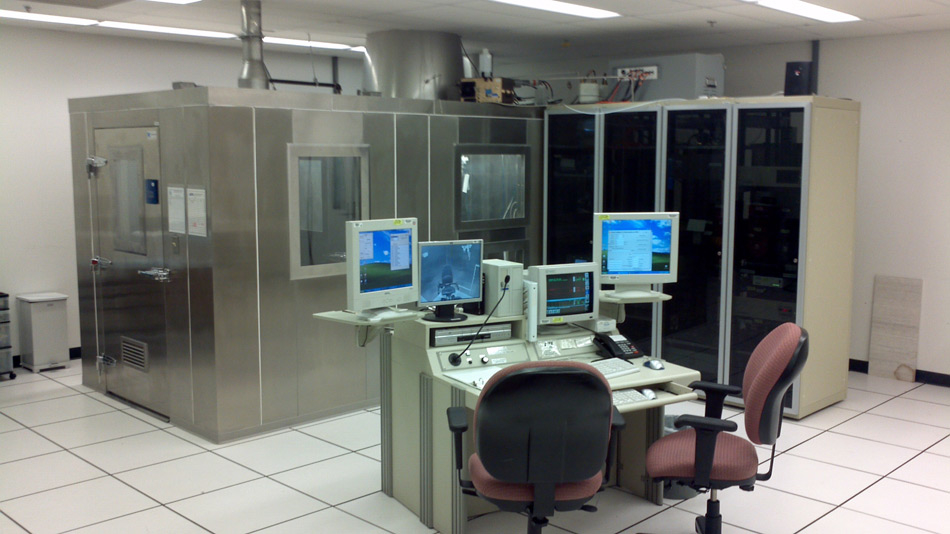
Figure 1
Diesel exhaust is collected from an idling truck parked outside the building (A). This is diluted with filtered air and introduced through piping into an exposure chamber (B).
In controlled human exposures frequently conducted in environmental health, human volunteers and patients are exposed either sequentially or at random to filtered air and a pollutant of interest. Exposures may employ DE, woodstove emissions, ozone, nitrogen oxides, sulfur oxides, carbon monoxide, resuspension of particles collected on filters, emission source and ambient particles (e.g., DEP and concentrated ambient particles), surrogate particles (e.g., carbon black), and combinations of these. Exposures can be performed in a concentration-related manner if subjects can be scheduled for repeated visits. Additionally, another advantage for the controlled experiment is an ability to perform time-dependent analyses; that is, the exposure can be turned on and off at specific intervals and the human response can then be assessed for pharmacokinetic parameters. Classical pharmacokinetic (PK) and physiologically based pharmacokinetic (PBPK) models for blood, breath, and urine biomarkers can be developed to explore biological variability due to metabolism and to provide a framework for exposure reconstruction for estimating previous exposures based on current biomarker measurements [13, 14]. Although such models could also be constructed from observational data in humans or from animal experiments by inferring various partition coefficients and rate constants, the controlled chamber experiment serves as the gold standard for assessing pharmacokinetic parameters.
Controlled human exposures at the United States Environmental Protection Agency employ DE generated from an idling 6-cylinder, 205 hp, 5.9L-displacement diesel engine (fig. 1A) in a vehicle located outside of the Human Studies Facility (Chapel Hill, North Carolina). Diesel fuel is purchased as a certified fuel and the same lot is used for the duration of the study. DE is introduced into the exposure chamber (1.83 m x 1.83 m x 2.44 m) after a 1:30 dilution with clean and humidified air to yield a feedback-regulated concentration of 100 to 300 μg/m3 (fig. 1B). In the exposure chamber, the volunteer alternates between sitting (15 minutes) and exercising (15 minutes) on a recumbent bicycle ergometer for two hours. The chamber temperature and humidity are controlled to approximately 22 ± 2oC and 40 ± 10% respectively. DEP is controlled in real time using a tapered element oscillating microbalance to measure the chamber concentration. The particle number concentration and the number size distribution are also measured. The particle concentration is verified by weighing filters obtained using a versatile air pollution sampler. In addition to these measures of particle size and mass, environmental measurements are numerous during the exposure (table 2). Health endpoints routinely obtained before and after controlled human exposures can include symptoms (with a computerised questionnaire), changes in physical examination, indices in blood, urine, breath condensate, exhaled breath, sputum, and bronchoalveolar lavage. Pulmonary function tests, heart rate variability, and Holter exams can also be repeatedly performed before and after the exposure. Bronchoscopy is frequently included to obtain evidence of an effect by the DE.
| Table 1: Strengths and weaknesses of controlled human exposure studies. | |
| Strengths | Weaknesses |
| Well-defined exposure concentrations. | Small subject numbers. |
| Clear durations of exposure. | Generally short term effects only. |
| Control of confounding exposures. | Usually cannot expose the most sensitive population. |
| Employment of a number of distinct health endpoints. | Lack of control for recent exposure. |
| Predetermined inclusion and exclusion criteria to possibly minimise variability. | |
| Inclusion of potentially susceptible populations (e.g., elderly, diabetics, and individuals with chronic obstructive pulmonary disease, asthma, and cardiovascular disease). | |
| Use of controlled exercise to increase dose and shorten exposure duration. | |
| Access to instrumentation not feasible to employ in field studies. | |
| Table 2: Common environmental measurements obtained during controlled human exposure to diesel exhaust. |
| Exposure chamber temperature Relative humidity Nitrogen dioxide Nitric oxide Sulfur dioxide Carbon monoxide Total hydrocarbons Particle mass concentration Particle number concentration Particle size |
Biomarkers are valuable tools for quantifying chemical exposures in epidemiological studies. Yet, applications of biomonitoring for DE exposure assessment have been limited because: 1) DE is a complex and dynamic mixture; 2) traditional exposure surrogates for DE (i.e., EC and PM) do not lend themselves to analysis in biological media; and 3) chemical constituents of DE that can be measured as biomarkers are often ubiquitous environmental pollutants. In a previously discussed controlled exposure study [15], a suite of volatile aromatic compounds, volatile and semivolatile PAHs, and particle-bound PAHs in chamber air was measured (both “control” air and 100 µg/m3 DEP plus combustion gases) and evaluated as potential DE exposure surrogates [16]. The goal of this analysis was to identify and critically evaluate promising biomarkers of DE exposure for use in epidemiological studies. First, airborne concentrations of individual analytes in DE were studied to identify prominent species. In general, volatile aromatic compounds (e.g., benzene, toluene, ethylbenzene, xylenes (BTEX) and styrene) stood out as the most abundant analytes, followed by volatile and semivolatile, PAHs (e.g., naphthalene and phenanthrene), and then particle-bound PAHs (e.g., chrysene and benzo(a)pyrene). Next, enrichment from the DE source was quantified using the ratio of analyte levels in DE to those in clean (purified and filtered) air. Ratios were largest for the particle-bound PAHs, followed by the volatile and semivolatile PAHs, and then the volatile aromatic compounds. The volatile and semivolatile PAHs, specifically napthalene and phenanthrene, were selected for additional evaluation since they were both abundant in DE (compared to the particulate PAHs) and highly differentiated with respect to clear air (compared to the volatile aromatic compounds). Pairwise comparisons showed significant correlations between naphthalene, phenanthrene and organic constituents of DE, namely total organics, total volatile and semivolatile PAHs, and total particle-bound PAHs.
Based on these results, a follow-up analysis was performed of PAH levels (including those of naphthalene and phenanthrene) in blood samples of these study subjects collected before, immediately after, and 24 hr after exposure to DE [16]. Results showed no effect of the controlled DE exposure on blood PAH levels. Thus, a 2 hour exposure to 100 µg/m3 DEP and associated combustion gases did not affect circulating levels of blood PAHs in the study subjects. This suggests that the controlled exposure levels were too low to elicit an increase above “background” levels stemming from environmental sources. There is also some possibility that PAH levels were either transiently elevated and then absent at 24 hours or not yet detectable at 24 hours. Future controlled studies of DE exposures may identify a threshold for distinguishing biomarkers of DE exposure from those of background/ambient sources and also optimal kinetics for sampling.
Gaseous and particulate components of air pollution initiate an inflammatory response in humans [17, 18]. This inflammation was considered essential in the mechanistic pathways of injury, including pulmonary and cardiovascular disease, following air pollution exposure. Since it is a constituent of ambient air pollution, it was anticipated that DE, and DEP, would contribute to a comparable set of biological effects. Epidemiologic investigation associated indices of both traffic-related air pollution and DE exposure with adverse human health outcomes including pulmonary inflammation, exacerbations of asthma, lung infections, and cardiovascular effects [19–23]. Accordingly, investigation using controlled human exposures to DE and DEP was undertaken.
Controlled human exposure to DE increased both neutrophils and B lymphocytes in bronchoalveolar lavage (BAL) of healthy volunteers 6 hours following a 1 hour exposure (with moderate intermittent exercise) to diluted whole DE (with mean DEP equivalent to 300 µg/m3) [24]. Lavage concentrations of histamine and fibronectin were similarly elevated. Biopsies confirmed an influx of neutrophils, mast cells, and T lymphocytes into the bronchial wall as well as elevated expression of adhesion molecules defining a lung inflammatory response. In addition to the lung, neutrophils were elevated in the peripheral blood supporting a systemic inflammation. Increased mRNA and protein expression of pro-inflammatory mediators were observed in cells resident in the lower respiratory tract [25]. These results were in agreement with an investigation (double-blind, randomised, and crossover) of 10 healthy, non-smoking volunteers exposed to resuspended DEP (mean DEP of 200 µg/m3) without the associated gases for 2 hours which demonstrated increased sputum neutrophils and sputum myeloperoxidase 4 hours later; however, there was no evidence of a systemic inflammation [26]. In addition to sputum induction and phlebotomy, endpoints in this study included serial spirometry and measurement of pulse, blood pressure, exhaled carbon monoxide, and methacholine reactivity at 0 to 4 and 24 hours after exposure. There were no changes in cardiovascular parameters or lung function following exposure to DEP but levels of exhaled CO as a marker of effect (i.e., heme oxygenase function) were increased and maximal at 1 hour. In a third investigation using controlled human exposure, fifteen healthy subjects exposed to DE (mean DEP of 270 µg/m3) showed evidence of lung inflammation on bronchoscopy [27]. There was an increased expression of p-selectin and vascular endothelial adhesion molecules in bronchial mucosal biopsies and elevated numbers of eosinophils in the BAL 6 hours after exposure. It was suggested that disparities between the results of this study [27] and that of prior investigation may reflect engine load [24]. Normal, healthy subjects exposed to DE (mean DEP of 200 µg/m3) for 2 hours at rest showed an activation of an inflammation pathway (proteosome activity) in peripheral white blood cells [28]. All of these studies support pro-inflammatory events in the lung following human exposure to DE or DEP.
There have also been controlled human exposure studies showing an absence of an effect of DE and DEP exposure on lung inflammation. Twenty-five healthy subjects exposed to diluted whole DE (100 µg/m3) demonstrated increased symptom prevalence and bronchoconstriction with increased values of airway resistance [29]. Spirometry failed to reveal any changes and there was no evidence of an inflammatory response on bronchoscopy with BAL 6 hours later. An increased concentration of reduced glutathione was evident in the airways. Among 15 non-smoking, healthy volunteers, exposure to DE (300 µg/m3) for 1 hour had no effect on bronchial expression of interleukin (IL)-10 and IL-8 in airway epithelial cells [30]. Results of a third study showed that 18 hours after exposure of healthy subjects to DE (100 µg/m3) for 2 hours, there was inflammation in the airways (increased neutrophil numbers, IL-8, and myeloperoxidase concentrations in bronchial lavage and elevated increased neutrophils and mast cells in bronchial mucosa) but not in the alveolar compartment [31].
As a result of epidemiologic investigation documenting a worsening of asthma following exposure to DE [19, 21–23, 32, 33], asthmatics were included in controlled human exposures. In one study, a group of 14 non-smoking, atopic asthmatics (treated with inhaled corticosteroids) were exposed to DE (300 µg/m3) for 1 hour [34]. While all participants were hyperresponsive at baseline, there was increased airway responsiveness (i.e., methacholine challenge) 24 hour following DE exposure. There was also an increase in airway resistance and elevated levels of sputum interleukin-6 but no changes in sputum levels of eosinophil cationic protein, myeloperoxidase and interleukin-8 and sputum differential cell counts. A second controlled human exposure to DE and DEP 108 µg/m3 for 2 hours similarly did not demonstrate an inflammatory response among asthmatics [35]. While healthy volunteers had airway neutrophilia, lymphocytosis, and lavage interleukin-6 and -8 concentrations 6 hours after the exposure, asthmatics showed no response. In a third study focused on asthmatic subjects, there was a lack of inflammation following exposure of subjects with mild to moderate disease to DE (mean DEP of 100 µg/m3) for 2 hours [36]. In contrast, healthy subjects showed increased neutrophil numbers, myeloperoxidase, and IL-6 in the bronchial wash and submucosal neutrophils on biopsy 18 hours after the same exposure.
These controlled human exposure studies support the conclusion that exposure of healthy subjects to DE and DEP is consistently pro-inflammatory only at higher concentrations with a threshold dose approximating 300 µg/m3. The pro-inflammatory effect shows considerable variability between the different exhausts and particles employed in investigation; disparities between these studies likely reflect disparities in the DE and DEP. Exposure concentration and duration as well as the timing of bronchoscopy need to be considered when inflammatory responses are evaluated. Additional factors including engine type, engine load, fuel characteristics, and driving mode may all modify the airway inflammatory responses to DE/DEP. There appears to be a lack of an inflammatory response to DE and DEP in the lungs of asthmatic individuals.
Controlled human exposure has also been employed in defining cardiovascular effects of DE and DEP. Exposure of 20 men (with a mean age 60 years) with prior myocardial infarction to DE (300 µg/m3) demonstrated worsening of exercise-induced ischemia [37]. Subjects had had a myocardial infarction more than 6 months prior to enrollment in this study, had been treated with primary angioplasty and stenting, and were receiving standard secondary preventive therapy. Men with unstable coronary disease, angina pectoris, history of arrhythmia, diabetes mellitus, uncontrolled hypertension, renal failure, and hepatic failure were excluded. Current smokers and men with asthma, significant occupational exposures, or an intercurrent illness were also not enrolled in this investigation. Endpoints included vascular studies (requiring brachial artery cannulation with a 27 gauge needle), plasma tissue plasminogen activator, plasminogen activator inhibitor-1, and C-reactive protein. Myocardial ischaemia was detected on electrocardiograph during exercise in all subjects. Relative to filtered air, there was a greater decrement of ST segment during exposure to diesel exhaust and this was interpreted as an increase in the exercise-induced ischemic burden. DE did not aggravate pre-existing vasomotor dysfunction measured as endothelium-dependent (acetylcholine) and endothelium-independent (nitroprusside) vasodilatation at 6 hours after exposure. Exposure to DE did reduce the acute bradykinin-induced release of endothelial tissue plasminogen activator. There were no differences in tissue plasminogen activator, plasminogen activator inhibitor-1, leukocyte and neutrophil counts, platelet counts, and C-reactive protein. These findings in patients with prior myocardial infarction contrasted those observed in healthy subjects in which 30 healthy men were exposed to DE (300 µg/m3) using the same exposure protocol (double-blind, randomised, crossover) [38]. DE attenuated increases in forearm blood flow induced by bradykinin, acetylcholine, and nitroprusside infusion measured 2 and 6 hours after exposure in these healthy volunteers. DE also suppressed the bradykinin-induced release in plasma tissue plasminogen activator 6 hours after exposure. Vascular dysfunction among 15 healthy volunteers 24 hours after exposure using the same protocol confirmed that DE reduced acetylcholine- and bradykinin-induced vasodilatation [39]. However, DE had no effects on endothelium-independent vasodilatation induced by either sodium nitroprusside or verapamil. DE did not attenuate bradykinin-induced increase in plasma tissue plasminogen activator.
In a fourth controlled human exposure study into the cardiovascular effects of DE, 19 healthy volunteers were exposed to DE for 1 hour [40]. Bilateral forearm blood flow and plasma fibrinolytic factors were assessed with venous occlusion plethysmography and blood sampling during intra-arterial infusion of acetylcholine, bradykinin, sodium nitroprusside, and verapamil. Compared with filtered air, DE decreased reduced vasodilatation and increased ex vivo thrombus formation. A particle trap reduced DEP number (from 150000 to 30000/cm3 to 30 to 300/cm3) and mass (320 ± 10 to 7.2 ± 2.0 μg/m3) and was associated with increased vasodilatation, reduced ex vivo thrombus formation, and an increase in tissue plasminogen activator release. The negative effects of DE on vasomotor function, endogenous fibrinolysis, and thrombus formation were precluded by reducing DEP concentration and number with a particle trap.
These controlled human exposure studies of the cardiovascular effects of DE and DEP do not allow broad conclusions. However, it appears that, comparable to other particle-associated exposures, DE can precipitate coronary artery responses. In addition, there is a relationship between DE and DEP exposure and vascular endpoints. Maximal effects on vasomotor function were detected at 2 and 6 hours after exposure, with some prolonged effects on acetylcholine responses at 24 hours. Effects on endogenous fibrinolysis were only detected at 6 hours. These effects may be associated with DEP rather than gaseous components of DE.
Findings derived from several studies support the concept that the biological effect of DE and DEP exposure is initiated by oxidative stress [41–47]. DEP produces reactive oxygen species in the lungs of animal models [48–51]. The oxidative stress associated with DE and DEP initiates a cascade of reactions culminating in inflammation. This includes phosphorylation-dependent cell signaling, an activation of the mitogen-activated protein (MAP) kinase cascade (ERK, p38, and Jun kinases), and release of pro-inflammatory mediators [47, 52–58]. Subsequently, the immediate response to DE and DEP is a pulmonary and systemic inflammation. Disparities between specific DEP in the capacity to induce oxidants and inflammation have been reported (e.g., forklift-generated DEP, NIST standard reference material 2975, and Japanese automobile-derived DEP show dissimilarities in biological effect). Those exact characteristics responsible for these differences are yet to be defined. However, they are likely the result of distinct physicochemical properties arising from different generation and collection conditions [59]. Generally, the capacity of DEP to produce both oxidative stress and a biological response approximates those of other combustion products such as wood stove particles [60–62].
The biological effect of DE and DEP is most frequently attributed to inflammation resulting from oxidant generation. The human controlled-exposure studies at elevated exposure levels (i.e., for DEP concentrations in the range of several hundred μg/m3) largely support the pro-inflammatory role of DE in humans. If prolonged, such inflammation has been associated with chronic sequellae including tissue fibrosis, cardiovascular disease, and increased risk for neoplasm [63–67]. It has been postulated that specific effects of particles can be the result of a systemic distribution of those with ultrafine size [68]. Cardiovascular effects of DEP are likely to be independent of such distribution for two reasons. First, it has been demonstrated that it is not necessary to invoke such systemic distribution to provoke a biological effect since other models of human inflammation associated with an increased risk for cardiovascular disease have never been shown to necessitate this (e.g., gingivitis affects an increased risk for cardiovascular disease without a requirement for septicemia) [69]. Second, there is conflicting data on whether translocation of insoluble particles from the lung into the bloodstream of any animal model actually occurs [70].
Investigation either directly demonstrates or supports a decrease in the inflammatory response to DE and DEP among those individuals with pre-existing disease (e.g., asthmatic, chronic obstructive pulmonary disease, and chronic heart disease) [37–39]. This was not anticipated as these are the same populations which appear to be at increased risk for particle-related morbidity and mortality in epidemiological studies [71, 72]. One explanation for this inconsistency is that individuals with chronic disease are frequently on medications which could alter the inflammatory response to DE and DEP exposure [37, 73–75]. Prominent among these medications would be the statins which affects the response to particle-associated exposures [76]. A second possibility to account for the diminished inflammatory effect in these patients is a selection bias for individuals with specific gene expression among those with chronic disease [76]. A final potential reason for disparities in the response between healthy volunteers and those with chronic illness to DE and DEP in controlled human exposures could be that disease process itself modifies the inflammatory reaction to DE and DEP exposure. In support of this, COPD patients demonstrate changes in patterns in particle deposition which can modify the response to DE and DEP exposure [77, 78]. Those with chronic inflammation may not have the ability to coordinate an acute inflammatory response to DE and DEP; this would be analogous to inflammation following infection [79]. It can be postulated that the acute inflammation benefits the individual and that the incapacity to fully initiate such inflammation in the chronically ill predicts a negative outcome.
The effects of DE have been investigated for their impact on human health more than any other emission source pollutant [80]. In the healthy subject, controlled human acute exposure to DE incites lung and systemic inflammation with a threshold concentration approximating 300 µg/m3. Systemic health consequences of DE exposure are considered consequent to the primary lung inflammation and include pro-thrombotic changes and cardiovascular disease. It appears that DEP is associated with some portion of the biological effect of DE. Future research can focus on potential effects of longer or repeated exposure, effects in sensitive individuals, the relative importance of DE components, and potential interactions between components and other pollutants.
1 Schauer JJ, Kleeman MJ, Cass GR, Simoneit BRT. Measurement of emissions from air pollution sources. 2. C-1 through C-30 organic compounds from medium duty diesel trucks. Environmental Science & Technology. 1999;33(10):1578–87.
2 Zielinska B, Sagebiel J, McDonald JD, Whitney K, Lawson DR. Emission rates and comparative chemical composition from selected in-use diesel and gasoline-fueled vehicles. J Air Waste Manag Assoc. 2004;54(9):1138–50.
3 Kittleson DB. Engines and nanoparticles: a review. Journal of Aerosol Science. 1998;29:575–88.
4 Gilman P. Health assessment document for diesel engine exhaust. US Environmental Protection Agency 2002.
5 Corfa E, Maury F, Segers P, Fresneau A, Albergel A. Short-range evaluation of air pollution near bus and railway stations. Sci Total Environ. 2004;334–335:223–30.
6 Cyrys J, Pitz M, Bischof W, Wichmann HE, Heinrich J. Relationship between indoor and outdoor levels of fine particle mass, particle number concentrations and black smoke under different ventilation conditions. J Expo Anal Environ Epidemiol. 2004;14(4):275–83.
7 Buzzard NA, Clark NN, Guffey SE. Investigation into pedestrian exposure to near-vehicle exhaust emissions. Environ Health. 2009;8:13.
8 Hesterberg TW, Lapin CA, Bunn WB. A comparison of emissions from vehicles fueled with diesel or compressed natural gas. Environ Sci Technol. 2008;42(17):6437–45.
9 Hesterberg TW, Long CM, Sax SN, Lapin CA, McClellan RO, Bunn WB, et al. Particulate matter in new technology diesel exhaust (NTDE) is quantitatively and qualitatively very different from that found in traditional diesel exhaust (TDE). J Air Waste Manag Assoc. 2011;61(9):894–913.
10 Kittleson DB, Watts WF, Johnson JP. On-road evaluation of two diesel exhaust aftertreatment devices. Aerosol Science. 2006;37:1140–51.
11 Utell MJ, Frampton MW. Toxicologic methods: controlled human exposures. Environ Health Perspect. 2000;108(Suppl 4):605–13.
12 McDonnell WF. Utility of controlled human exposure studies for assessing the health effects of complex mixtures and indoor air pollutants. Environ Health Perspect. 1993;101(Suppl 4):199–203.
13 Pleil JD, Kim D, Prah JD, Rappaport SM. Exposure reconstruction for reducing uncertainty in risk assessment: example using MTBE biomarkers and a simple pharmacokinetic model. Biomarkers. 2007;12(4):331–48.
14 Kim D, Andersen ME, Pleil JD, Nylander-French LA, Prah JD. Refined PBPK model of aggregate exposure to methyl tertiary-butyl ether. Toxicol Lett. 2007;169(3):222–35.
15 Sobus JR, Pleil JD, Madden MC, Funk WE, Hubbard HF, Rappaport SM. Identification of surrogate measures of diesel exhaust exposure in a controlled chamber study. Environ Sci Technol. 2008;42(23):8822–8.
16 Pleil JD, Sheldon LS. Adapting concepts from systems biology to develop systems exposure event networks for exposure science research. Biomarkers. 2011;16(2):99–105.
17 Bromberg PA, Koren HS. Ozone-induced human respiratory dysfunction and disease. Toxicol Lett. 1995;82-83:307–16.
18 Ghio AJ, Kim C, Devlin RB. Concentrated ambient air particles induce mild pulmonary inflammation in healthy human volunteers. Am J Respir Crit Care Med. 2000;162(3 Pt 1):981–8.
19 Escamilla-Nunez MC, Barraza-Villarreal A, Hernandez-Cadena L, Moreno-Macias H, Ramirez-Aguilar M, Sienra-Monge JJ, et al. Traffic-related air pollution and respiratory symptoms among asthmatic children, resident in Mexico City: the EVA cohort study. Respir Res. 2008;9:74.
20 Ostro B, Roth L, Malig B, Marty M. The effects of fine particle components on respiratory hospital admissions in children. Environ Health Perspect. 2009;117(3):475–80.
21 Mar TF, Koenig JQ, Primomo J. Associations between asthma emergency visits and particulate matter sources, including diesel emissions from stationary generators in Tacoma, Washington. Inhal Toxicol. 2010;22(6):445–8.
22 Patel MM, Chillrud SN, Correa JC, Hazi Y, Feinberg M, Kc D, et al. Traffic-related particulate matter and acute respiratory symptoms among New York City area adolescents. Environ Health Perspect. 2010;118(9):1338–43.
23 Spira-Cohen A, Chen LC, Kendall M, Lall R, Thurston GD. Personal exposures to traffic-related air pollution and acute respiratory health among Bronx schoolchildren with asthma. Environ Health Perspect. 2011;119(4):559–65.
24 Salvi S, Blomberg A, Rudell B, Kelly F, Sandstrom T, Holgate ST, et al. Acute inflammatory responses in the airways and peripheral blood after short-term exposure to diesel exhaust in healthy human volunteers. Am J Respir Crit Care Med. 1999;159(3):702–9.
25 Salvi SS, Nordenhall C, Blomberg A, Rudell B, Pourazar J, Kelly FJ, et al. Acute exposure to diesel exhaust increases IL-8 and GRO-alpha production in healthy human airways. Am J Respir Crit Care Med. 2000;161(2 Pt 1):550–7.
26 Nightingale JA, Maggs R, Cullinan P, Donnelly LE, Rogers DF, Kinnersley R, et al. Airway inflammation after controlled exposure to diesel exhaust particulates. Am J Respir Crit Care Med. 2000;162(1):161–6.
27 Sehlstedt M, Behndig AF, Boman C, Blomberg A, Sandstrom T, Pourazar J. Airway inflammatory response to diesel exhaust generated at urban cycle running conditions. Inhal Toxicol. 2010;22(14):1144–50.
28 Kipen HM, Gandhi S, Rich DQ, Ohman-Strickland P, Laumbach R, Fan ZH, et al. Acute decreases in proteasome pathway activity after inhalation of fresh diesel exhaust or secondary organic aerosol. Environ Health Perspect. 2011;119(5):658–63.
29 Mudway IS, Stenfors N, Duggan ST, Roxborough H, Zielinski H, Marklund SL, et al. An in vitro and in vivo investigation of the effects of diesel exhaust on human airway lining fluid antioxidants. Arch Biochem Biophys. 2004;423(1):200–12.
30 Pourazar J, Frew AJ, Blomberg A, Helleday R, Kelly FJ, Wilson S, et al. Diesel exhaust exposure enhances the expression of IL-13 in the bronchial epithelium of healthy subjects. Respir Med. 2004;98(9):821–5.
31 Behndig AF, Mudway IS, Brown JL, Stenfors N, Helleday R, Duggan ST, et al. Airway antioxidant and inflammatory responses to diesel exhaust exposure in healthy humans. Eur Respir J. 2006;27(2):359–65.
32 Hoppin JA, Umbach DM, London SJ, Alavanja MC, Sandler DP. Diesel exhaust, solvents, and other occupational exposures as risk factors for wheeze among farmers. Am J Respir Crit Care Med. 2004;169(12):1308–13.
33 Green RS, Basu R, Malig B, Broadwin R, Kim JJ, Ostro B. The effect of temperature on hospital admissions in nine California counties. Int J Public Health. 2010;55(2):113–21.
34 Nordenhall C, Pourazar J, Ledin MC, Levin JO, Sandstrom T, Adelroth E. Diesel exhaust enhances airway responsiveness in asthmatic subjects. Eur Respir J. 2001;17(5):909–15.
35 Stenfors N, Nordenhall C, Salvi SS, Mudway I, Soderberg M, Blomberg A, et al. Different airway inflammatory responses in asthmatic and healthy humans exposed to diesel. Eur Respir J. 2004;23(1):82–6.
36 Behndig AF, Larsson N, Brown JL, Stenfors N, Helleday R, Duggan ST, et al. Proinflammatory doses of diesel exhaust in healthy subjects fail to elicit equivalent or augmented airway inflammation in subjects with asthma. Thorax. 2011;66(1):12–9.
37 Mills NL, Tornqvist H, Gonzalez MC, Vink E, Robinson SD, Soderberg S, et al. Ischemic and thrombotic effects of dilute diesel-exhaust inhalation in men with coronary heart disease. N Engl J Med. 2007;357(11):1075–82.
38 Mills NL, Tornqvist H, Robinson SD, Gonzalez M, Darnley K, MacNee W, et al. Diesel exhaust inhalation causes vascular dysfunction and impaired endogenous fibrinolysis. Circulation. 2005;112(25):3930–6.
39 Tornqvist H, Mills NL, Gonzalez M, Miller MR, Robinson SD, Megson IL, et al. Persistent endothelial dysfunction in humans after diesel exhaust inhalation. Am J Respir Crit Care Med. 2007;176(4):395–400.
40 Lucking AJ, Lundback M, Barath SL, Mills NL, Sidhu MK, Langrish JP, et al. Particle traps prevent adverse vascular and prothrombotic effects of diesel engine exhaust inhalation in men. Circulation. 2011;123(16):1721–8.
41 Jaspers I, Ciencewicki JM, Zhang W, Brighton LE, Carson JL, Beck MA, et al. Diesel exhaust enhances influenza virus infections in respiratory epithelial cells. Toxicol Sci. 2005;85(2):990–1002.
42 Alaghmand M, Blough NV. Source-dependent variation in hydroxyl radical production by airborne particulate matter. Environ Sci Technol. 2007;41(7):2364–70.
43 Cherng TW, Paffett ML, Jackson-Weaver O, Campen MJ, Walker BR, Kanagy NL. Mechanisms of diesel-induced endothelial nitric oxide synthase dysfunction in coronary arterioles. Environ Health Perspect. 2011;119(1):98–103.
44 DiStefano E, Eiguren-Fernandez A, Delfino RJ, Sioutas C, Froines JR, Cho AK. Determination of metal-based hydroxyl radical generating capacity of ambient and diesel exhaust particles. Inhal Toxicol. 2009;21(9):731–8.
45 Kumagai Y, Arimoto T, Shinyashiki M, Shimojo N, Nakai Y, Yoshikawa T, et al. Generation of reactive oxygen species during interaction of diesel exhaust particle components with NADPH-cytochrome P450 reductase and involvement of the bioactivation in the DNA damage. Free Radic Biol Med. 1997;22(3):479–87.
46 Li R, Ning Z, Majumdar R, Cui J, Takabe W, Jen N, et al. Ultrafine particles from diesel vehicle emissions at different driving cycles induce differential vascular pro-inflammatory responses: implication of chemical components and NF-kappaB signaling. Part Fibre Toxicol. 2010;7:6.
47 Marano F, Boland S, Bonvallot V, Baulig A, Baeza-Squiban A. Human airway epithelial cells in culture for studying the molecular mechanisms of the inflammatory response triggered by diesel exhaust particles. Cell Biol Toxicol. 2002;18(5):315–20.
48 Ichinose T, Furuyama A, Sagai M. Biological effects of diesel exhaust particles (DEP). II. Acute toxicity of DEP introduced into lung by intratracheal instillation. Toxicology. 1995;99(3):153–67.
49 Lim HB, Ichinose T, Miyabara Y, Takano H, Kumagai Y, Shimojyo N, et al. Involvement of superoxide and nitric oxide on airway inflammation and hyperresponsiveness induced by diesel exhaust particles in mice. Free Radic Biol Med. 1998;25(6):635–44.
50 Sagai M, Furuyama A, Ichinose T. Biological effects of diesel exhaust particles (DEP). III. Pathogenesis of asthma like symptoms in mice. Free Radic Biol Med. 1996;21(2):199–209.
51 Sagai M, Saito H, Ichinose T, Kodama M, Mori Y. Biological effects of diesel exhaust particles. I. In vitro production of superoxide and in vivo toxicity in mouse. Free Radic Biol Med. 1993;14(1):37–47.
52 Amara N, Bachoual R, Desmard M, Golda S, Guichard C, Lanone S, et al. Diesel exhaust particles induce matrix metalloprotease-1 in human lung epithelial cells via a NADP(H) oxidase/NOX4 redox-dependent mechanism. Am J Physiol Lung Cell Mol Physiol. 2007;293(1):L170–81.
53 Li R, Ning Z, Cui J, Khalsa B, Ai L, Takabe W, et al. Ultrafine particles from diesel engines induce vascular oxidative stress via JNK activation. Free Radic Biol Med. 2009;46(6):775–82.
54 Li N, Kim S, Wang M, Froines J, Sioutas C, Nel A. Use of a stratified oxidative stress model to study the biological effects of ambient concentrated and diesel exhaust particulate matter. Inhal Toxicol. 2002;14(5):459–86.
55 Su B, Karin M. Mitogen-activated protein kinase cascades and regulation of gene expression. Curr Opin Immunol. 1996;8(3):402–11.
56 Xiao GG, Wang M, Li N, Loo JA, Nel AE. Use of proteomics to demonstrate a hierarchical oxidative stress response to diesel exhaust particle chemicals in a macrophage cell line. J Biol Chem. 2003;278(50):50781–90.
57 Li N, Alam J, Venkatesan MI, Eiguren-Fernandez A, Schmitz D, Di Stefano E, et al. Nrf2 is a key transcription factor that regulates antioxidant defense in macrophages and epithelial cells: protecting against the proinflammatory and oxidizing effects of diesel exhaust chemicals. J Immunol. 2004;173(5):3467–81.
58 Pourazar J, Mudway IS, Samet JM, Helleday R, Blomberg A, Wilson SJ, et al. Diesel exhaust activates redox-sensitive transcription factors and kinases in human airways. Am J Physiol Lung Cell Mol Physiol. 2005;289(5):L724–30.
59 Singh P, DeMarini DM, Dick CA, Tabor DG, Ryan JV, Linak WP, et al. Sample characterization of automobile and forklift diesel exhaust particles and comparative pulmonary toxicity in mice. Environ Health Perspect. 2004;112(8):820–5.
60 Stoeger T, Reinhard C, Takenaka S, Schroeppel A, Karg E, Ritter B, et al. Instillation of six different ultrafine carbon particles indicates a surface area threshold dose for acute lung inflammation in mice. Environ Health Perspect. 2006;114(3):328–33.
61 Seagrave J, McDonald JD, Reed MD, Seilkop SK, Mauderly JL. Responses to subchronic inhalation of low concentrations of diesel exhaust and hardwood smoke measured in rat bronchoalveolar lavage fluid. Inhal Toxicol. 2005;17(12):657–70.
62 Seagrave J, McDonald JD, Gigliotti AP, Nikula KJ, Seilkop SK, Gurevich M, et al. Mutagenicity and in vivo toxicity of combined particulate and semivolatile organic fractions of gasoline and diesel engine emissions. Toxicol Sci. 2002;70(2):212–26.
63 Khansari N, Shakiba Y, Mahmoudi M. Chronic inflammation and oxidative stress as a major cause of age-related diseases and cancer. Recent Pat Inflamm Allergy Drug Discov. 2009;3(1):73–80.
64 Lakhan SE, Kirchgessner A, Hofer M. Inflammatory mechanisms in ischemic stroke: therapeutic approaches. J Transl Med. 2009;7:97.
65 Hulsmans M, Holvoet P. The vicious circle between oxidative stress and inflammation in atherosclerosis. J Cell Mol Med. 2010;14(1-2):70–8.
66 Brevetti G, Giugliano G, Brevetti L, Hiatt WR. Inflammation in peripheral artery disease. Circulation. 2010;122(18):1862–75.
67 Bringardner BD, Baran CP, Eubank TD, Marsh CB. The role of inflammation in the pathogenesis of idiopathic pulmonary fibrosis. Antioxid Redox Signal. 2008;10(2):287–301.
68 Terzano C, Di Stefano F, Conti V, Graziani E, Petroianni A. Air pollution ultrafine particles: toxicity beyond the lung. Eur Rev Med Pharmacol Sci. 2010;14(10):809–21.
69 Humphrey LL, Fu R, Buckley DI, Freeman M, Helfand M. Periodontal disease and coronary heart disease incidence: a systematic review and meta-analysis. J Gen Intern Med. 2008;23(12):2079–86.
70 Mills NL, Amin N, Robinson SD, Anand A, Davies J, Patel D, et al. Do inhaled carbon nanoparticles translocate directly into the circulation in humans? Am J Respir Crit Care Med. 2006;173(4):426–31.
71 Dockery DW, Pope CA, 3rd, Xu X, Spengler JD, Ware JH, Fay ME, et al. An association between air pollution and mortality in six U.S. cities. N Engl J Med. 1993;329(24):1753–9.
72 Samet JM, Dominici F, Curriero FC, Coursac I, Zeger SL. Fine particulate air pollution and mortality in 20 U.S. cities, 1987–1994. N Engl J Med. 2000;343(24):1742–9.
73 Wheeler A, Zanobetti A, Gold DR, Schwartz J, Stone P, Suh HH. The relationship between ambient air pollution and heart rate variability differs for individuals with heart and pulmonary disease. Environ Health Perspect. 2006;114(4):560–6.
74 Routledge HC, Manney S, Harrison RM, Ayres JG, Townend JN. Effect of inhaled sulphur dioxide and carbon particles on heart rate variability and markers of inflammation and coagulation in human subjects. Heart. 2006;92(2):220–7.
75 Gong H, Jr., Linn WS, Clark KW, Anderson KR, Geller MD, Sioutas C. Respiratory responses to exposures with fine particulates and nitrogen dioxide in the elderly with and without COPD. Inhal Toxicol. 2005;17(3):123–32.
76 Schwartz J, Park SK, O’Neill MS, Vokonas PS, Sparrow D, Weiss S, et al. Glutathione-S-transferase M1, obesity, statins, and autonomic effects of particles: gene-by-drug-by-environment interaction. Am J Respir Crit Care Med. 2005;172(12):1529–33.
77 Kim CS, Kang TC. Comparative measurement of lung deposition of inhaled fine particles in normal subjects and patients with obstructive airway disease. Am J Respir Crit Care Med. 1997;155(3):899–905.
78 Volterrani M, Scalvini S, Mazzuero G, Lanfranchi P, Colombo R, Clark AL, et al. Decreased heart rate variability in patients with chronic obstructive pulmonary disease. Chest. 1994;106(5):1432–7.
79 Rieber N, Hector A, Kuijpers T, Roos D, Hartl D. Current concepts of hyperinflammation in chronic granulomatous disease. Clin Dev Immunol. 2012;2012:252460.
80 Mauderly JL. Diesel emissions: is more health research still needed? Toxicol Sci. 2001;62(1):6–9.
Disclaimer: This report has been reviewed by the National Health and Environmental Effects Research Laboratory, United States Environmental Protection Agency and approved for publication. Approval does not signify that the contents necessarily reflect the views and policies of the Agency nor does mention of trade names or commercial products constitute endorsement or recommendation for use.
Funding / potential competing interests: No financial support and no other potential conflict of interest relevant to this article was reported.